Rana Vivek, Srivastava Nikhil, Kaushik Noopur, Sharma Vrinda, Panthri Prerna, Niranjan Madan Mohan
Department of Pediatric and Preventive Dentistry, Subharti Dental College and Hospital, Swami Vivekanand Subharti University, Meerut, Uttar Pradesh, India.
Int J Clin Pediatr Dent. 2019 Jan-Feb;12(1):64-67. doi: 10.5005/jp-journals-10005-1575.
Odontomas generally appear as small, solitary, or multiple radio-opaque lesions found on routine radiographic examinations. Traditionally, odontomas have been classified as benign odontogenic tumors and are subdivided into complex or compound odontomas morphologically. Frequently, they interfere with the eruption of the teeth.
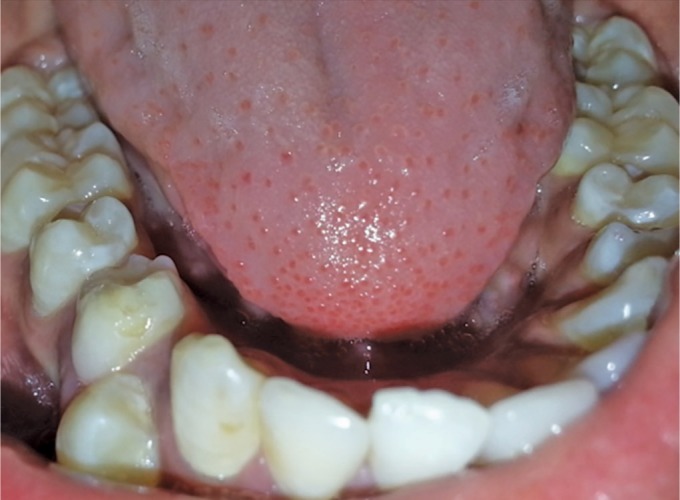
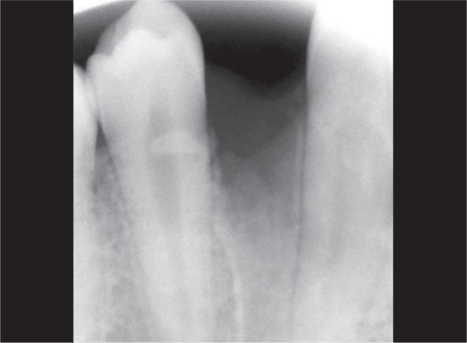
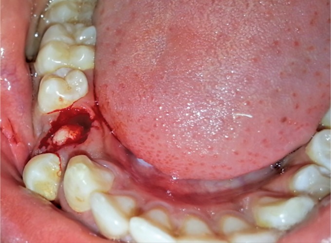
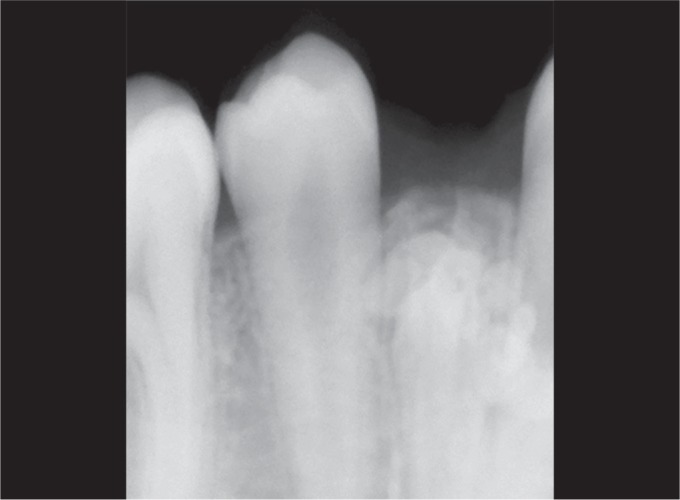
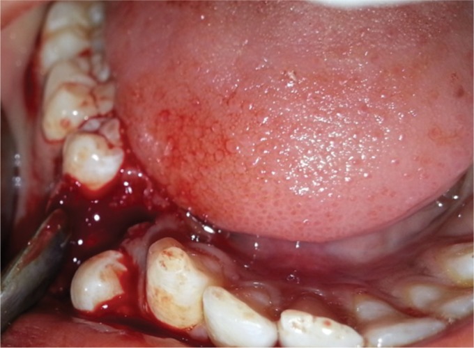
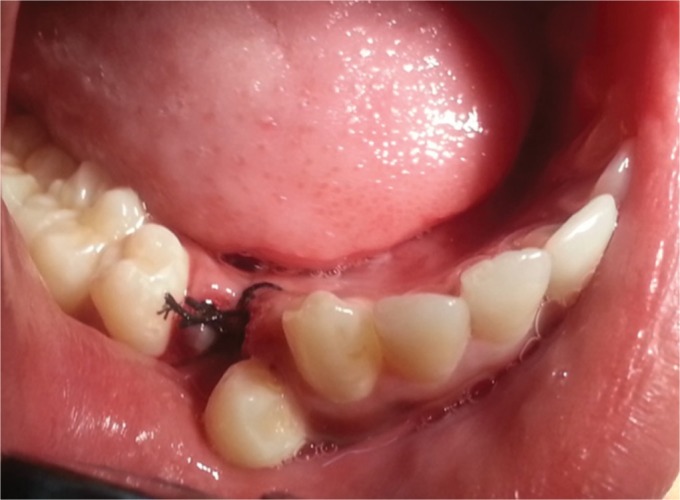
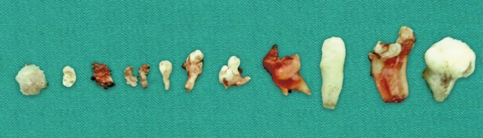
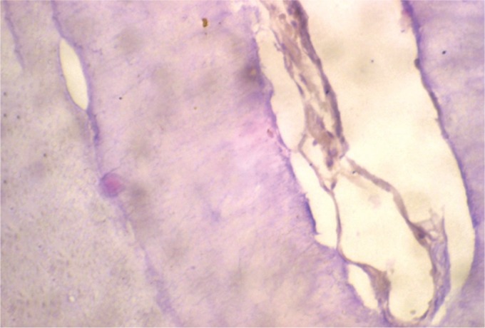
This paper describes the case of a compound odontoma in a 10-year-old boy diagnosed after extraction of the retained right primary mandibular first molar in the radiograph. A surgical excision was performed and the histopathological examination revealed a compound odontome.
Early diagnosis of odontomas and complete removal ensures better prognosis.
Rana V, Srivastava N, Compound Odontome: A Case Report. Int J Clin Pediatr Dent 2019;12(1):64-67.
牙瘤通常表现为在常规影像学检查中发现的小的、孤立的或多发的不透射线病变。传统上,牙瘤被归类为良性牙源性肿瘤,在形态学上可细分为复合性或组合性牙瘤。它们经常会干扰牙齿的萌出。
本文描述了一名10岁男孩的复合性牙瘤病例,该病例是在X线片显示拔除保留的右下第一乳磨牙后确诊的。进行了手术切除,组织病理学检查显示为复合性牙瘤。
牙瘤的早期诊断和彻底切除可确保更好的预后。
Rana V, Srivastava N, 复合性牙瘤:一例报告。《国际临床儿科牙科学杂志》2019年;12(1):64 - 67。