• 文献检索
  • 文档翻译
  • 深度研究
  • 学术资讯
  • Suppr Zotero 插件Zotero 插件
  • 邀请有礼
  • 套餐&价格
  • 历史记录
应用&插件
Suppr Zotero 插件Zotero 插件浏览器插件Mac 客户端Windows 客户端微信小程序
定价
高级版会员购买积分包购买API积分包
服务
文献检索文档翻译深度研究API 文档MCP 服务
关于我们
关于 Suppr公司介绍联系我们用户协议隐私条款
关注我们

Suppr 超能文献

核心技术专利:CN118964589B侵权必究
粤ICP备2023148730 号-1Suppr @ 2026

文献检索

告别复杂PubMed语法,用中文像聊天一样搜索,搜遍4000万医学文献。AI智能推荐,让科研检索更轻松。

立即免费搜索

文件翻译

保留排版,准确专业,支持PDF/Word/PPT等文件格式,支持 12+语言互译。

免费翻译文档

深度研究

AI帮你快速写综述,25分钟生成高质量综述,智能提取关键信息,辅助科研写作。

立即免费体验

脑出血猪模型中血肿周围组织的系列代谢评估

Serial Metabolic Evaluation of Perihematomal Tissues in the Intracerebral Hemorrhage Pig Model.

作者信息

Haque Muhammad E, Gabr Refaat E, George Sarah D, Zhao Xiurong, Boren Seth B, Zhang Xu, Ting Shun-Ming, Sun Gunghua, Hasan Khader M, Savitz Sean, Aronowski Jaroslaw

机构信息

Institute for Stroke and Cerebrovascular Diseases, McGovern Medical School, The University of Texas Health Science Center at Houston, Houston, TX, United States.

Diagnostic and Interventional Imaging, McGovern Medical School, The University of Texas Health Science Center at Houston, Houston, TX, United States.

出版信息

Front Neurosci. 2019 Aug 21;13:888. doi: 10.3389/fnins.2019.00888. eCollection 2019.

DOI:10.3389/fnins.2019.00888
PMID:31496934
原文链接:https://pmc.ncbi.nlm.nih.gov/articles/PMC6712426/
Abstract

PURPOSE

Perihematomal edema (PHE) occurs in patients with intracerebral hemorrhage (ICH) and is often used as surrogate of secondary brain injury. PHE resolves over time, but little is known about the functional integrity of the tissues that recover from edema. In a pig ICH model, we aimed to assess metabolic integrity of perihematoma tissues by using non-invasive magnetic resonance spectroscopy (MRS).

MATERIALS AND METHODS

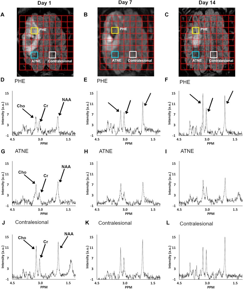
Fourteen male Yorkshire pigs with an average age of 8 weeks were intracerebrally injected with autologous blood to produce ICH. Proton MRS data were obtained at 1, 7, and 14 days after ICH using a whole-body 3.0T MRI system. Point-resolved spectroscopy (PRESS)-localized 2D chemical shift imaging (CSI) was acquired. The concentration of -Acetylaspartate (NAA), Choline (Cho), and Creatine (Cr) were measured within the area of PHE, tissues adjacent to the injury with no or negligible edema (ATNE), and contralesional brain tissue. A linear mixed model was used to analyze the evolution of metabolites in perihematomal tissues, with -value < 0.05 indicating statistical significance.

RESULTS

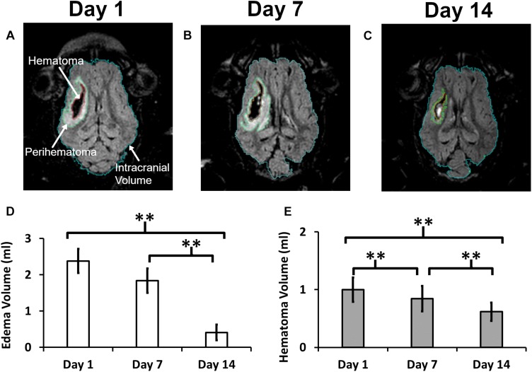
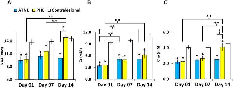
The perihematoma volume gradually decreased from 2.38 ± 1.23 ml to 0.41 ± 0.780 ml ( < 0.001) over 2 weeks. Significant ( 0.001) reductions in NAA, Cr, and Cho concentrations were found in the PHE and ATNE regions compared to the contralesional hemisphere at day 1 and 7 after ICH. All three metabolites were significantly ( < 0.001) restored in the PHE tissue on day 14, but remained persistently low in the ATNE area, and unaltered in the contralesional voxel.

CONCLUSION

This study highlights the potential of MRS to probe salvageable tissues within the perihematoma in the sub-acute phase of ICH. Altered metabolites within the PHE and ATNE regions in addition to edema and hematoma volumes were explored as possible markers for tissue recovery. Perihematomal tissue with PHE demonstrated a more reversible injury compared to the tissue adjacent to the injury without edema, suggesting a potentially beneficial role of edema.

摘要

目的

脑出血(ICH)患者会出现血肿周围水肿(PHE),其常被用作继发性脑损伤的替代指标。PHE会随着时间消退,但对于从水肿中恢复的组织的功能完整性知之甚少。在猪ICH模型中,我们旨在通过使用非侵入性磁共振波谱(MRS)评估血肿周围组织的代谢完整性。

材料与方法

14只平均年龄为8周的雄性约克夏猪经脑内注射自体血以产生ICH。在ICH后1天、7天和14天,使用全身3.0T MRI系统获取质子MRS数据。采集点分辨波谱(PRESS)定位的二维化学位移成像(CSI)。在PHE区域、无或仅有可忽略水肿的损伤邻近组织(ATNE)以及对侧脑组织内测量N-乙酰天门冬氨酸(NAA)、胆碱(Cho)和肌酸(Cr)的浓度。使用线性混合模型分析血肿周围组织中代谢物的演变,P值<0.05表示具有统计学意义。

结果

在2周内,血肿周围体积从2.38±1.23 ml逐渐减小至0.41±0.780 ml(P<0.001)。与对侧半球相比,在ICH后第1天和第7天,PHE和ATNE区域的NAA、Cr和Cho浓度显著降低(P<0.001)。在第14天,PHE组织中所有三种代谢物均显著恢复(P<0.001),但在ATNE区域仍持续较低,而在对侧体素中未改变。

结论

本研究突出了MRS在ICH亚急性期探测血肿周围可挽救组织的潜力。除了水肿和血肿体积外,还探索了PHE和ATNE区域内代谢物的改变作为组织恢复的可能标志物。与无水肿的损伤邻近组织相比,伴有PHE的血肿周围组织表现出更可逆的损伤,提示水肿可能具有有益作用。

https://cdn.ncbi.nlm.nih.gov/pmc/blobs/042b/6712426/070a1abec8fd/fnins-13-00888-g003.jpg
https://cdn.ncbi.nlm.nih.gov/pmc/blobs/042b/6712426/994f8f2bfc15/fnins-13-00888-g001.jpg
https://cdn.ncbi.nlm.nih.gov/pmc/blobs/042b/6712426/95bcc412f5ba/fnins-13-00888-g002.jpg
https://cdn.ncbi.nlm.nih.gov/pmc/blobs/042b/6712426/070a1abec8fd/fnins-13-00888-g003.jpg
https://cdn.ncbi.nlm.nih.gov/pmc/blobs/042b/6712426/994f8f2bfc15/fnins-13-00888-g001.jpg
https://cdn.ncbi.nlm.nih.gov/pmc/blobs/042b/6712426/95bcc412f5ba/fnins-13-00888-g002.jpg
https://cdn.ncbi.nlm.nih.gov/pmc/blobs/042b/6712426/070a1abec8fd/fnins-13-00888-g003.jpg

相似文献

1
Serial Metabolic Evaluation of Perihematomal Tissues in the Intracerebral Hemorrhage Pig Model.脑出血猪模型中血肿周围组织的系列代谢评估
Front Neurosci. 2019 Aug 21;13:888. doi: 10.3389/fnins.2019.00888. eCollection 2019.
2
Cytotoxic Edema and Adverse Clinical Outcomes in Patients with Intracerebral Hemorrhage.细胞毒性水肿与脑出血患者的不良临床转归。
Neurocrit Care. 2023 Apr;38(2):414-421. doi: 10.1007/s12028-022-01603-2. Epub 2022 Sep 30.
3
Longitudinal, Quantitative, Multimodal MRI Evaluation of Patients With Intracerebral Hemorrhage Over the First Year.脑出血患者第一年的纵向、定量、多模态磁共振成像评估
Front Neurol. 2021 Nov 30;12:764718. doi: 10.3389/fneur.2021.764718. eCollection 2021.
4
Clinical implications of Peri-hematomal edema microperfusion fraction in intracerebral hemorrhage intravoxel incoherent motion imaging - A pilot study.血肿周围水肿微灌注分数在脑出血体素内不相干运动成像中的临床意义——一项初步研究。
J Stroke Cerebrovasc Dis. 2023 Nov;32(11):107375. doi: 10.1016/j.jstrokecerebrovasdis.2023.107375. Epub 2023 Sep 20.
5
Sex differences in perihematomal edema volume and outcome after intracerebral hemorrhage.脑出血后血肿周围水肿体积及预后的性别差异
medRxiv. 2023 Sep 29:2023.09.28.23296302. doi: 10.1101/2023.09.28.23296302.
6
Serial Cerebral Metabolic Changes in Patients With Ischemic Stroke Treated With Autologous Bone Marrow Derived Mononuclear Cells.接受自体骨髓来源单核细胞治疗的缺血性中风患者的系列脑代谢变化
Front Neurol. 2019 Feb 25;10:141. doi: 10.3389/fneur.2019.00141. eCollection 2019.
7
Association of serum occludin levels and perihematomal edema volumes in intracranial hemorrhage patients.脑出血患者血清紧密连接蛋白水平与血肿周围水肿体积的相关性。
CNS Neurosci Ther. 2024 Mar;30(3):e14450. doi: 10.1111/cns.14450. Epub 2023 Sep 18.
8
Quantitative assessment on blood-brain barrier permeability of acute spontaneous intracerebral hemorrhage in basal ganglia: a CT perfusion study.基底节区急性自发性脑出血血脑屏障通透性的定量评估:一项CT灌注研究
Neuroradiology. 2017 Jul;59(7):677-684. doi: 10.1007/s00234-017-1852-9. Epub 2017 Jun 3.
9
Perihematomal edema in primary intracerebral hemorrhage is plasma derived.原发性脑出血中的血肿周围水肿源自血浆。
Stroke. 2004 Aug;35(8):1879-85. doi: 10.1161/01.STR.0000131807.54742.1a. Epub 2004 Jun 3.
10
Perihematomal edema: Implications for intracerebral hemorrhage research and therapeutic advances.血肿周围水肿:对脑出血研究和治疗进展的影响。
J Neurosci Res. 2020 Jan;98(1):212-218. doi: 10.1002/jnr.24372. Epub 2018 Dec 21.

引用本文的文献

1
Molecular, Pathological, Clinical, and Therapeutic Aspects of Perihematomal Edema in Different Stages of Intracerebral Hemorrhage.血肿不同阶段的血肿周围水肿的分子、病理、临床和治疗方面。
Oxid Med Cell Longev. 2022 Sep 17;2022:3948921. doi: 10.1155/2022/3948921. eCollection 2022.
2
Defining Delayed Perihematomal Edema Expansion in Intracerebral Hemorrhage: Segmentation, Time Course, Risk Factors and Clinical Outcome.定义脑出血后血肿周围水肿延迟扩大:分割、时间进程、危险因素和临床转归。
Front Immunol. 2022 May 9;13:911207. doi: 10.3389/fimmu.2022.911207. eCollection 2022.
3
Changes in Cerebral Blood Flow and Diffusion-Weighted Imaging Lesions After Intracerebral Hemorrhage.

本文引用的文献

1
Inflammatory Markers and Severity of Intracerebral Hemorrhage.炎症标志物与脑出血的严重程度
Cureus. 2018 Oct 31;10(10):e3529. doi: 10.7759/cureus.3529.
2
Perihematomal edema: Implications for intracerebral hemorrhage research and therapeutic advances.血肿周围水肿:对脑出血研究和治疗进展的影响。
J Neurosci Res. 2020 Jan;98(1):212-218. doi: 10.1002/jnr.24372. Epub 2018 Dec 21.
3
Early hematoma retraction in intracerebral hemorrhage is uncommon and does not predict outcome.脑出血早期血肿回缩并不常见,也不能预测结果。
脑出血后脑血流和弥散加权成像病变的变化。
Transl Stroke Res. 2022 Oct;13(5):686-706. doi: 10.1007/s12975-022-00998-6. Epub 2022 Mar 19.
4
Perihematomal Edema After Intracerebral Hemorrhage: An Update on Pathogenesis, Risk Factors, and Therapeutic Advances.脑出血后血肿周围水肿:发病机制、危险因素和治疗进展的更新。
Front Immunol. 2021 Oct 19;12:740632. doi: 10.3389/fimmu.2021.740632. eCollection 2021.
5
Neuroimaging of Acute Intracerebral Hemorrhage.急性脑出血的神经影像学
J Clin Med. 2021 Mar 5;10(5):1086. doi: 10.3390/jcm10051086.
6
Relevance of Porcine Stroke Models to Bridge the Gap from Pre-Clinical Findings to Clinical Implementation.猪卒中模型与从临床前发现到临床实施的相关性。
Int J Mol Sci. 2020 Sep 8;21(18):6568. doi: 10.3390/ijms21186568.
7
Dl-3-n-Butylphthalide promotes neovascularization and neurological recovery in a rat model of intracerebral hemorrhage.二正丁基苯酞促进脑出血大鼠模型的血管新生和神经功能恢复。
BMC Neurosci. 2020 May 29;21(1):24. doi: 10.1186/s12868-020-00575-3.
PLoS One. 2018 Oct 9;13(10):e0205436. doi: 10.1371/journal.pone.0205436. eCollection 2018.
4
Absolute risk and predictors of the growth of acute spontaneous intracerebral haemorrhage: a systematic review and meta-analysis of individual patient data.急性自发性脑出血增长的绝对风险和预测因素:一项个体患者数据的系统回顾和荟萃分析。
Lancet Neurol. 2018 Oct;17(10):885-894. doi: 10.1016/S1474-4422(18)30253-9. Epub 2018 Aug 14.
5
Early Hemolysis Within Human Intracerebral Hematomas: an MRI Study.颅内血肿早期溶血:一项 MRI 研究。
Transl Stroke Res. 2019 Feb;10(1):52-56. doi: 10.1007/s12975-018-0630-2. Epub 2018 May 15.
6
Peak perihemorrhagic edema correlates with functional outcome in intracerebral hemorrhage.脑内出血时,出血周围水肿峰值与功能结局相关。
Neurology. 2018 Mar 20;90(12):e1005-e1012. doi: 10.1212/WNL.0000000000005167. Epub 2018 Feb 16.
7
Serial quantitative neuroimaging of iron in the intracerebral hemorrhage pig model.脑出血猪模型中铁的连续定量神经影像学研究。
J Cereb Blood Flow Metab. 2018 Mar;38(3):375-381. doi: 10.1177/0271678X17751548. Epub 2018 Jan 2.
8
Neutrophil polarization by IL-27 as a therapeutic target for intracerebral hemorrhage.白细胞介素-27介导的中性粒细胞极化作为脑出血的治疗靶点
Nat Commun. 2017 Sep 19;8(1):602. doi: 10.1038/s41467-017-00770-7.
9
Programmed Cell Death after Intracerebral Hemorrhage.脑出血后的细胞程序性死亡。
Curr Neuropharmacol. 2018;16(9):1267-1281. doi: 10.2174/1570159X15666170602112851.
10
Secondary Hematoma Expansion and Perihemorrhagic Edema after Intracerebral Hemorrhage: From Bench Work to Practical Aspects.脑出血后的继发性血肿扩大及出血周围水肿:从基础研究到实际应用
Front Neurol. 2017 Apr 7;8:74. doi: 10.3389/fneur.2017.00074. eCollection 2017.