Behrens Martin, Husmann Florian, Mau-Moeller Anett, Schlegel Jenny, Reuter Eva-Maria, Zschorlich Volker R
Institute of Sport Science, University of Rostock, Rostock, Germany.
Centre for Sensorimotor Performance, School of Human Movement and Nutrition Sciences, The University of Queensland, Brisbane, QLD, Australia.
Front Bioeng Biotechnol. 2019 Aug 21;7:181. doi: 10.3389/fbioe.2019.00181. eCollection 2019.
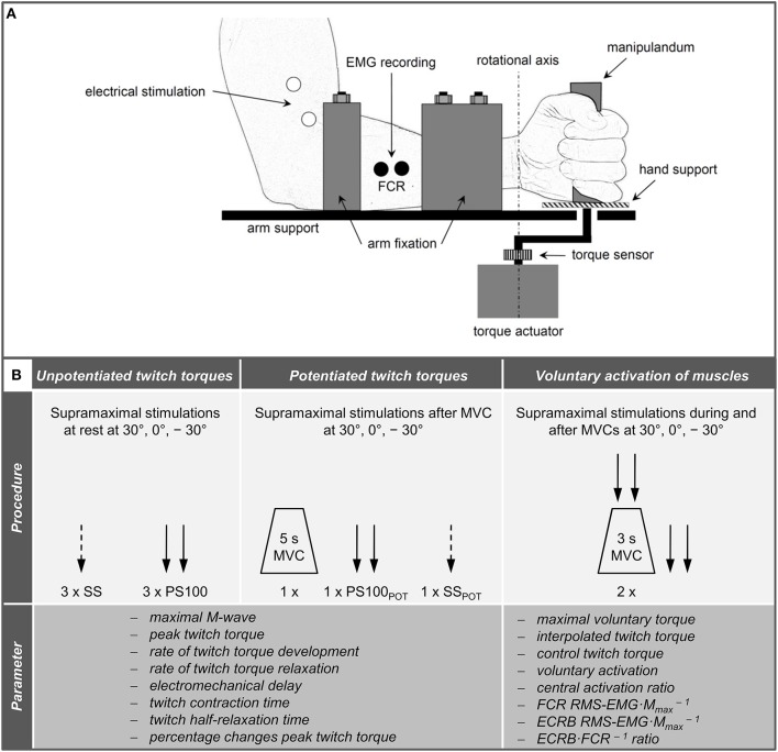
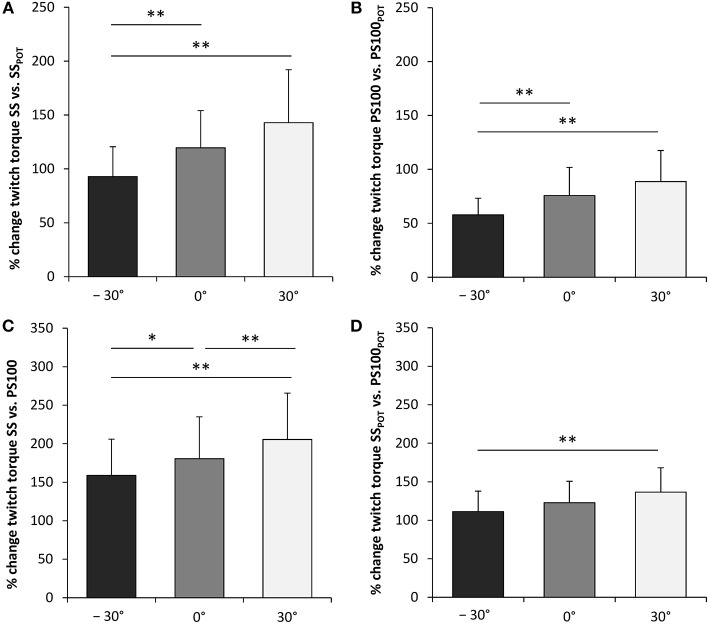
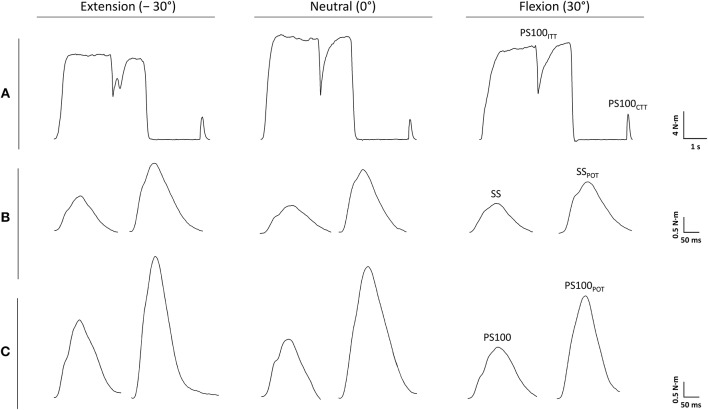
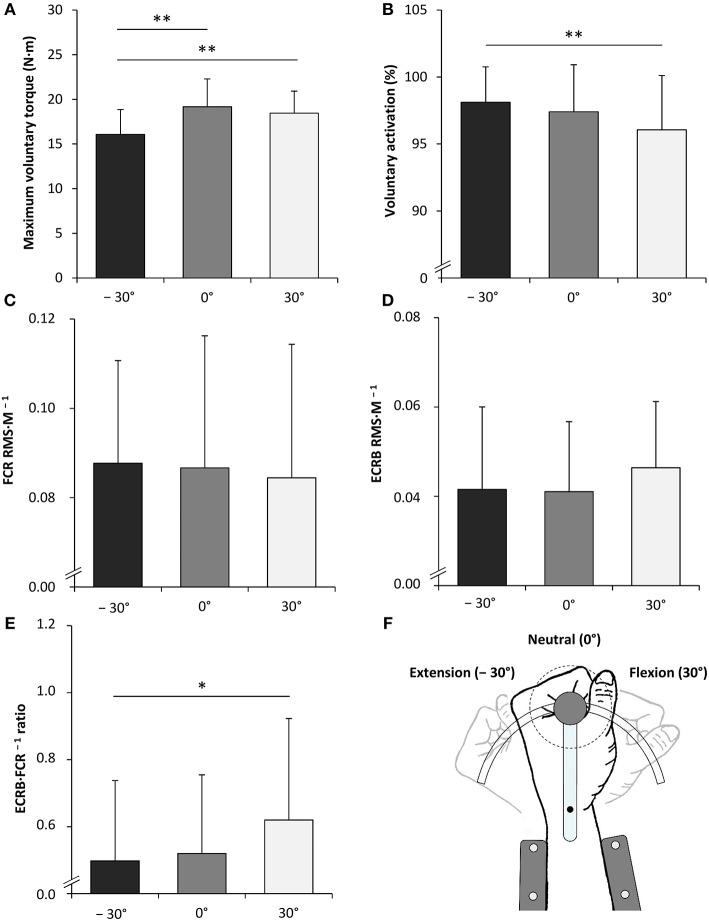
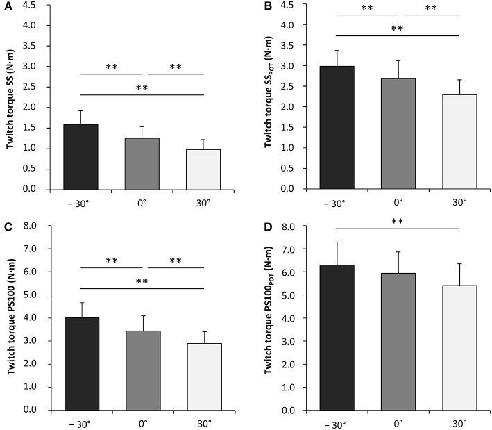
The joint angle dependence of voluntary activation and twitch properties has been investigated for several human skeletal muscles. However, although they play a key role for hand function and possess a unique neural control compared to muscles surrounding other joint complexes, little is known about the wrist flexors innervated by the median nerve. Therefore, isometric voluntary and electrically evoked contractions of the wrist flexors were analyzed at three wrist joint angles (extension: -30°, neutral: 0°, flexion: 30°) to quantify the joint angle dependence of (i) voluntary activation (assessed via peripheral nerve stimulation and electromyography [EMG]), (ii) unpotentiated twitch torques, and (iii) potentiated twitch torques. Maximum voluntary torque was lower in extension compared to neutral and flexion. Although voluntary activation was generally high, data indicate that voluntary activation of the wrist flexors innervated by the median nerve was lower and the antagonist·agonist EMG ratio was higher with the wrist joint in flexion compared to extension. Peak twitch torque, rate of twitch torque development, and twitch half-relaxation time increased, whereas electromechanical delay decreased from flexion to extension for the unpotentiated twitch torques. Activity-induced potentiation partly abolished these differences and was higher in short than long wrist flexors. Different angle-dependent excitatory and inhibitory inputs to spinal and supraspinal centers might be responsible for the altered activation of the investigated wrist muscles. Potential mechanisms were discussed and might have operated conjointly to increase stiffness of the flexed wrist joint. Differences in twitch torque properties were probably related to angle-dependent alterations in series elastic properties, actin-myosin interaction, Ca sensitivity, and phosphorylation of myosin regulatory light chains. The results of the present study provide valuable information about the contribution of neural and muscular properties to changes in strength capabilities of the wrist flexors at different wrist joint angles. These data could help to understand normal wrist function, which is a first step in determining mechanisms underlying musculoskeletal disorders and in giving recommendations for the restoration of musculoskeletal function after traumatic or overuse injuries.
已经对多块人体骨骼肌的随意激活和抽搐特性的关节角度依赖性进行了研究。然而,尽管它们对手部功能起着关键作用,并且与围绕其他关节复合体的肌肉相比具有独特的神经控制,但对于由正中神经支配的腕屈肌却知之甚少。因此,在三个腕关节角度(伸展:-30°,中立:0°,屈曲:30°)下分析了腕屈肌的等长随意收缩和电诱发收缩,以量化以下方面的关节角度依赖性:(i)随意激活(通过外周神经刺激和肌电图[EMG]评估),(ii)未增强的抽搐扭矩,以及(iii)增强的抽搐扭矩。与中立和屈曲相比,伸展时的最大随意扭矩较低。尽管随意激活通常较高,但数据表明,与伸展相比,腕关节屈曲时由正中神经支配的腕屈肌的随意激活较低,拮抗肌·主动肌EMG比率较高。对于未增强的抽搐扭矩,从屈曲到伸展,抽搐扭矩峰值、抽搐扭矩发展速率和抽搐半松弛时间增加,而机电延迟减少。活动诱导的增强部分消除了这些差异,并且在短腕屈肌中比长腕屈肌中更高。对脊髓和脊髓上中枢的不同角度依赖性兴奋性和抑制性输入可能是所研究的腕部肌肉激活改变的原因。讨论了潜在机制,这些机制可能共同作用以增加屈曲腕关节的刚度。抽搐扭矩特性的差异可能与串联弹性特性、肌动蛋白-肌球蛋白相互作用、钙敏感性和肌球蛋白调节轻链磷酸化的角度依赖性改变有关。本研究结果提供了有关神经和肌肉特性对不同腕关节角度下腕屈肌力量能力变化的贡献的有价值信息。这些数据有助于理解正常的腕部功能,这是确定肌肉骨骼疾病潜在机制以及为创伤或过度使用损伤后肌肉骨骼功能恢复提供建议的第一步。