Departamento de Neurobiología Celular y Molecular, Instituto de Neurobiología, Campus Juriquilla, Universidad Nacional Autónoma de México, Querétaro, Qro., 76230, Mexico.
Department of Physiology, University of Alberta, Edmonton, AB T6G 2H7, Canada.
Int J Mol Sci. 2019 Sep 10;20(18):4433. doi: 10.3390/ijms20184433.
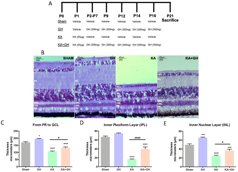
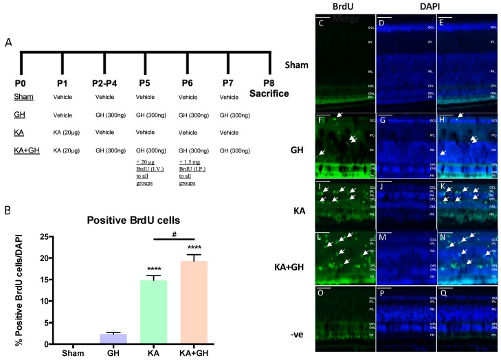
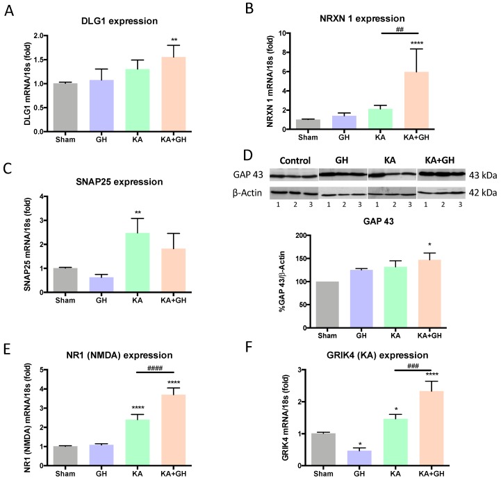
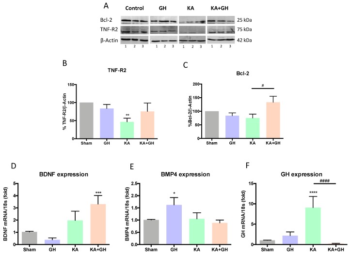
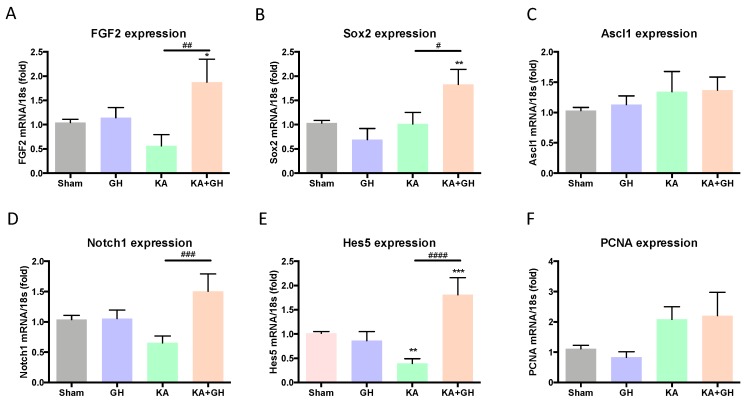
In addition to its role as an endocrine messenger, growth hormone (GH) also acts as a neurotrophic factor in the central nervous system (CNS), whose effects are involved in neuroprotection, axonal growth, and synaptogenic modulation. An increasing amount of clinical evidence shows a beneficial effect of GH treatment in patients with brain trauma, stroke, spinal cord injury, impaired cognitive function, and neurodegenerative processes. In response to injury, Müller cells transdifferentiate into neural progenitors and proliferate, which constitutes an early regenerative process in the chicken retina. In this work, we studied the long-term protective effect of GH after causing severe excitotoxic damage in the retina. Thus, an acute neural injury was induced via the intravitreal injection of kainic acid (KA, 20 µg), which was followed by chronic administration of GH (10 injections [300 ng] over 21 days). Damage provoked a severe disruption of several retinal layers. However, in KA-damaged retinas treated with GH, we observed a significant restoration of the inner plexiform layer (IPL, 2.4-fold) and inner nuclear layer (INL, 1.5-fold) thickness and a general improvement of the retinal structure. In addition, we also observed an increase in the expression of several genes involved in important regenerative pathways, including: synaptogenic markers (DLG1, NRXN1, GAP43); glutamate receptor subunits (NR1 and GRIK4); pro-survival factors (BDNF, Bcl-2 and TNF-R2); and Notch signaling proteins (Notch1 and Hes5). Interestingly, Müller cell transdifferentiation markers (Sox2 and FGF2) were upregulated by this long-term chronic GH treatment. These results are consistent with a significant increase in the number of BrdU-positive cells observed in the KA-damaged retina, which was induced by GH administration. Our data suggest that GH is able to facilitate the early proliferative response of the injured retina and enhance the regeneration of neurite interconnections.
除了作为内分泌信使的作用外,生长激素 (GH) 还在中枢神经系统 (CNS) 中充当神经营养因子,其作用涉及神经保护、轴突生长和突触发生调节。越来越多的临床证据表明 GH 治疗对脑外伤、中风、脊髓损伤、认知功能障碍和神经退行性过程患者具有有益的影响。在受伤后,Müller 细胞向神经祖细胞分化并增殖,这构成了鸡视网膜早期再生过程的一部分。在这项工作中,我们研究了 GH 在视网膜发生严重兴奋性损伤后的长期保护作用。因此,通过在玻璃体内注射海人酸 (KA,20 µg) 诱导急性神经损伤,随后用 GH 进行慢性治疗 (21 天内注射 10 次[300 ng])。损伤导致视网膜的几个层严重中断。然而,在接受 GH 治疗的 KA 损伤视网膜中,我们观察到内丛状层 (IPL,2.4 倍) 和内核层 (INL,1.5 倍) 厚度的显著恢复,以及视网膜结构的普遍改善。此外,我们还观察到一些参与重要再生途径的基因表达增加,包括:突触形成标记物 (DLG1、NRXN1、GAP43);谷氨酸受体亚基 (NR1 和 GRIK4);生存因子 (BDNF、Bcl-2 和 TNF-R2);和 Notch 信号蛋白 (Notch1 和 Hes5)。有趣的是,这种长期慢性 GH 治疗上调了 Müller 细胞分化标记物 (Sox2 和 FGF2)。这些结果与 GH 给药诱导的 KA 损伤视网膜中观察到的 BrdU 阳性细胞数量的显著增加一致。我们的数据表明,GH 能够促进受伤视网膜的早期增殖反应,并增强神经突连接的再生。