Hamlyn Centre, Institute of Global Health Innovation, Imperial College London, London, UK.
Department of Surgery and Cancer, Imperial College London, London, UK.
Sci Rep. 2022 May 21;12(1):8607. doi: 10.1038/s41598-022-12504-x.
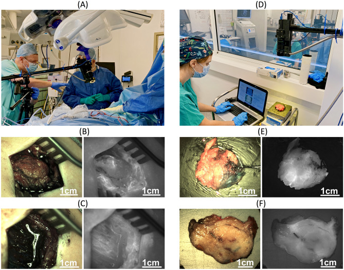
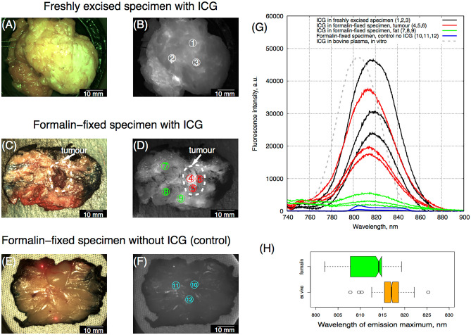
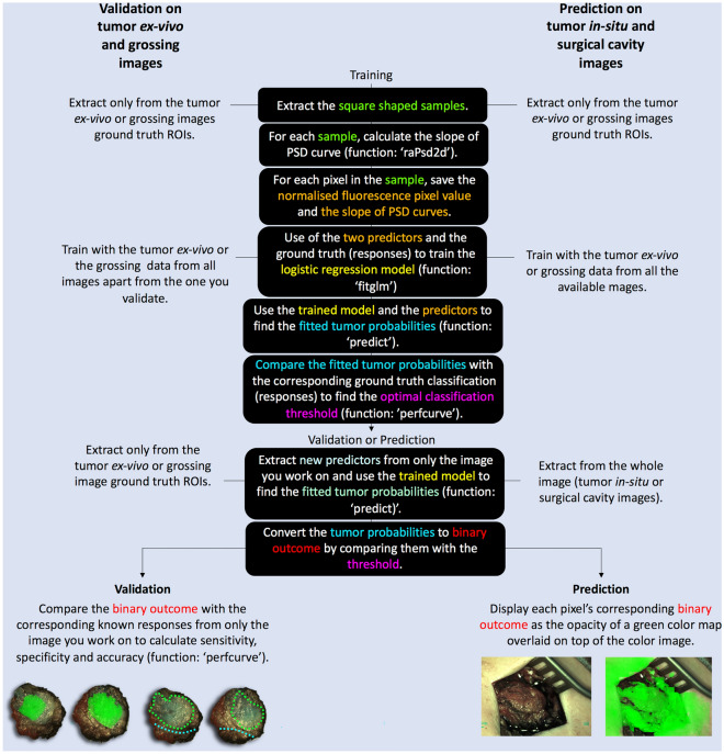
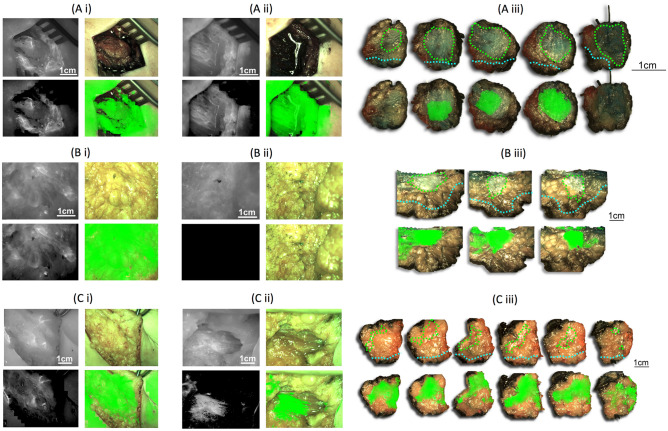
Re-operation due to disease being inadvertently close to the resection margin is a major challenge in breast conserving surgery (BCS). Indocyanine green (ICG) fluorescence imaging could be used to visualize the tumor boundaries and help surgeons resect disease more efficiently. In this work, ICG fluorescence and color images were acquired with a custom-built camera system from 40 patients treated with BCS. Images were acquired from the tumor in-situ, surgical cavity post-excision, freshly excised tumor and histopathology tumour grossing. Fluorescence image intensity and texture were used as individual or combined predictors in both logistic regression (LR) and support vector machine models to predict the tumor extent. ICG fluorescence spectra in formalin-fixed histopathology grossing tumor were acquired and analyzed. Our results showed that ICG remains in the tissue after formalin fixation. Therefore, tissue imaging could be validated in freshly excised and in formalin-fixed grossing tumor. The trained LR model with combined fluorescence intensity (pixel values) and texture (slope of power spectral density curve) identified the tumor's extent in the grossing images with pixel-level resolution and sensitivity, specificity of 0.75 ± 0.3, 0.89 ± 0.2.This model was applied on tumor in-situ and surgical cavity (post-excision) images to predict tumor presence.
由于疾病无意中接近切除边缘,导致再次手术是保乳手术 (BCS) 的主要挑战。吲哚菁绿 (ICG) 荧光成像是一种可以用来可视化肿瘤边界并帮助外科医生更有效地切除肿瘤的方法。在这项工作中,我们使用定制的相机系统从 40 名接受 BCS 治疗的患者中获取了 ICG 荧光和彩色图像。图像是从肿瘤原位、切除后的手术腔、新鲜切除的肿瘤和组织病理学大体肿瘤中获取的。荧光图像强度和纹理被用作逻辑回归 (LR) 和支持向量机模型的单个或组合预测因子,以预测肿瘤范围。我们还获取和分析了福尔马林固定组织病理学大体肿瘤中的 ICG 荧光光谱。结果表明,ICG 在福尔马林固定后仍保留在组织中。因此,组织成像可以在新鲜切除和福尔马林固定大体肿瘤中得到验证。使用联合荧光强度(像素值)和纹理(功率谱密度曲线斜率)的训练后的 LR 模型以像素级分辨率和 0.75±0.3、0.89±0.2 的灵敏度和特异性识别了大体图像中的肿瘤范围。该模型应用于肿瘤原位和手术腔(切除后)图像,以预测肿瘤的存在。