• 文献检索
  • 文档翻译
  • 深度研究
  • 学术资讯
  • Suppr Zotero 插件Zotero 插件
  • 邀请有礼
  • 套餐&价格
  • 历史记录
应用&插件
Suppr Zotero 插件Zotero 插件浏览器插件Mac 客户端Windows 客户端微信小程序
定价
高级版会员购买积分包购买API积分包
服务
文献检索文档翻译深度研究API 文档MCP 服务
关于我们
关于 Suppr公司介绍联系我们用户协议隐私条款
关注我们

Suppr 超能文献

核心技术专利:CN118964589B侵权必究
粤ICP备2023148730 号-1Suppr @ 2026

文献检索

告别复杂PubMed语法,用中文像聊天一样搜索,搜遍4000万医学文献。AI智能推荐,让科研检索更轻松。

立即免费搜索

文件翻译

保留排版,准确专业,支持PDF/Word/PPT等文件格式,支持 12+语言互译。

免费翻译文档

深度研究

AI帮你快速写综述,25分钟生成高质量综述,智能提取关键信息,辅助科研写作。

立即免费体验

基于 MRI 的影像组学对软组织肉瘤进行肿瘤分级。

Tumor grading of soft tissue sarcomas using MRI-based radiomics.

机构信息

Department of Radiation Oncology, Klinikum Rechts der Isar, School of Medicine, Technical University of Munich (TUM), Ismaninger Straße 22, 81675 Munich, Germany; Institute of Radiation Medicine (IRM), Department of Radiation Sciences (DRS), Helmholtz Zentrum München, Ingolstaedter Landstrasse 1, 85764 Neuherberg, Germany; Deutsches Konsortium für Translationale Krebsforschung (DKTK), Partner Site Munich, Germany.

Department of Radiation Oncology, University of Washington, 1959 NE Pacific St, Box 356043, Seattle, WA 98195, United States of America.

出版信息

EBioMedicine. 2019 Oct;48:332-340. doi: 10.1016/j.ebiom.2019.08.059. Epub 2019 Sep 12.

DOI:10.1016/j.ebiom.2019.08.059
PMID:31522983
原文链接:
https://pmc.ncbi.nlm.nih.gov/articles/PMC6838361/
Abstract

BACKGROUND

Treatment decisions for multimodal therapy in soft tissue sarcoma (STS) patients greatly depend on the differentiation between low-grade and high-grade tumors. We developed MRI-based radiomics grading models for the differentiation between low-grade (G1) and high-grade (G2/G3) STS.

METHODS

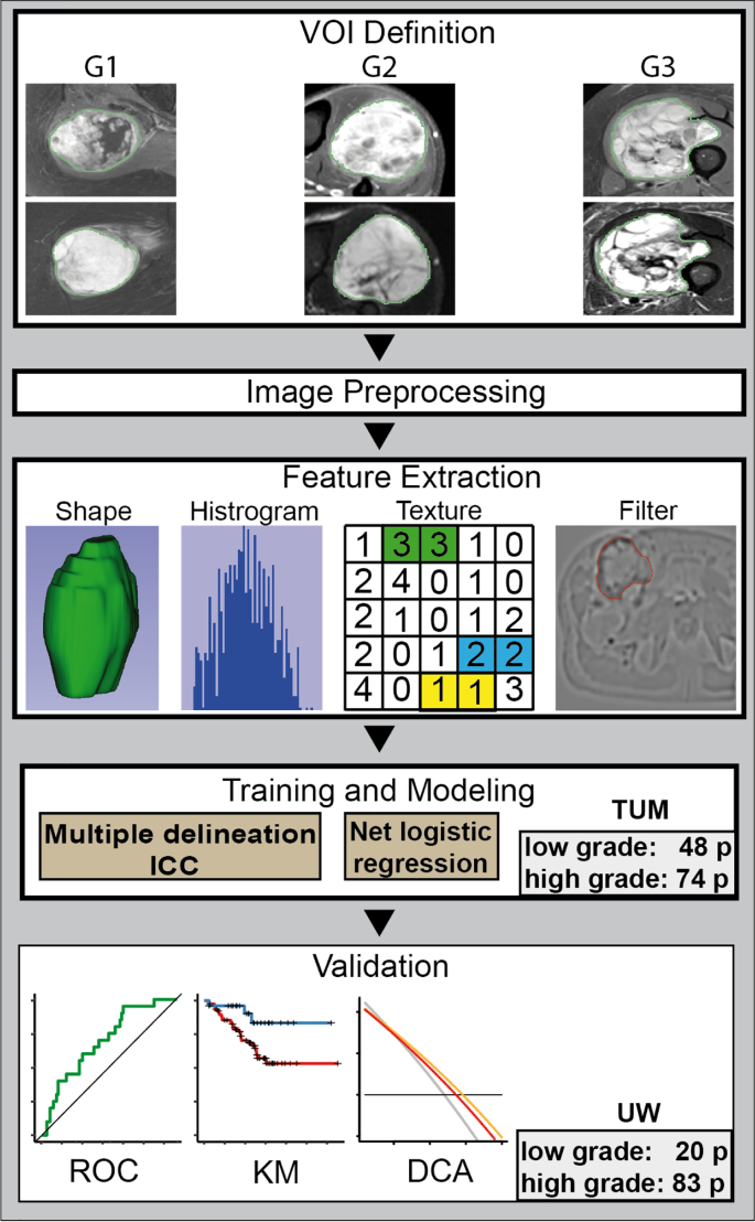
The study was registered at ClinicalTrials.gov (number NCT03798795). Contrast-enhanced T1-weighted fat saturated (T1FSGd), fat-saturated T2-weighted (T2FS) MRI sequences, and tumor grading following the French Federation of Cancer Centers Sarcoma Group obtained from pre-therapeutic biopsies were gathered from two independent retrospective patient cohorts. Volumes of interest were manually segmented. After preprocessing, 1394 radiomics features were extracted from each sequence. Features unstable in 21 independent multiple-segmentations were excluded. Least absolute shrinkage and selection operator models were developed using nested cross-validation on a training patient cohort (122 patients). The influence of ComBatHarmonization was assessed for correction of batch effects.

FINDINGS

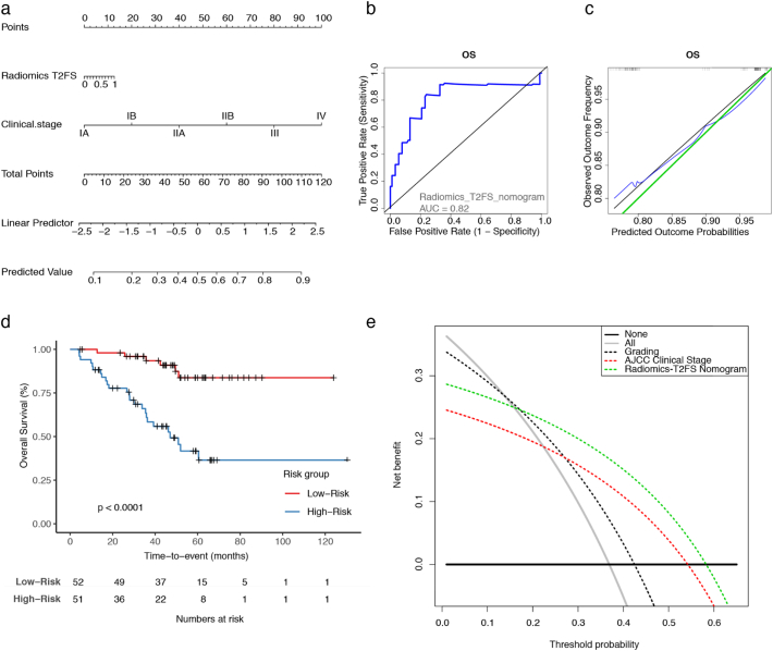
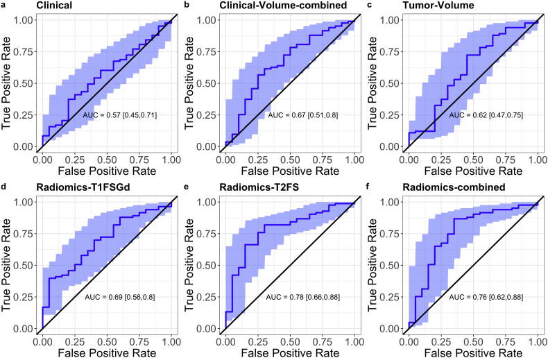
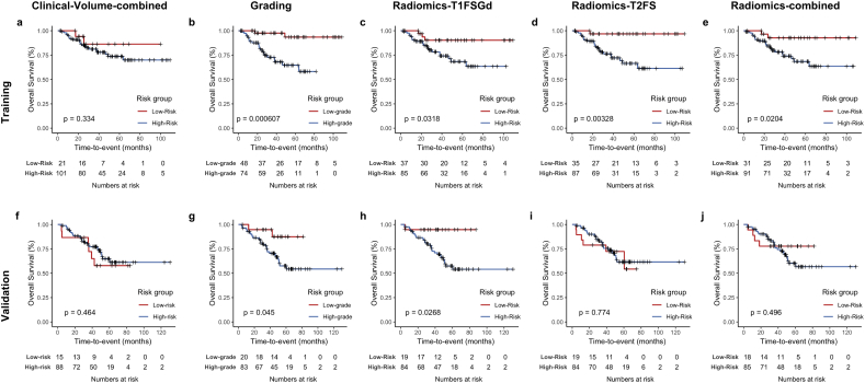
Three radiomic models based on T2FS, T1FSGd and a combined model achieved predictive performances with an area under the receiver operator characteristic curve (AUC) of 0.78, 0.69, and 0.76 on the independent validation set (103 patients), respectively. The T2FS-based model showed the best reproducibility. The radiomics model involving T1FSGd-based features achieved significant patient stratification. Combining the T2FS radiomic model into a nomogram with clinical staging improved prognostic performance and the clinical net benefit above clinical staging alone.

INTERPRETATION

MRI-based radiomics tumor grading models effectively classify low-grade and high-grade soft tissue sarcomas. FUND: The authors received support by the medical faculty of the Technical University of Munich and the German Cancer Consortium.

摘要

背景

软组织肉瘤(STS)多模态治疗的决策在很大程度上取决于低级别和高级别肿瘤的区分。我们开发了基于 MRI 的放射组学分级模型,用于区分低级别(G1)和高级别(G2/G3)STS。

方法

该研究在 ClinicalTrials.gov 注册(编号 NCT03798795)。从两个独立的回顾性患者队列中收集了术前活检获得的对比增强 T1 加权脂肪饱和(T1FSGd)、脂肪饱和 T2 加权(T2FS)MRI 序列和肿瘤分级,遵循法国癌症中心肉瘤组的标准。手动分割感兴趣体积。预处理后,从每个序列中提取 1394 个放射组学特征。从 21 个独立的多分割中提取不稳定的特征被排除。使用嵌套交叉验证在训练患者队列(122 例)上开发最小绝对收缩和选择算子模型。使用 ComBatHarmonization 评估了校正批次效应的影响。

发现

基于 T2FS、T1FSGd 和联合模型的三个放射组学模型在独立验证集(103 例)上的预测性能分别为接收器操作特征曲线(AUC)下面积为 0.78、0.69 和 0.76。基于 T2FS 的模型显示出最佳的可重复性。基于 T1FSGd 特征的放射组学模型实现了显著的患者分层。将 T1FSGd 基于特征的放射组学模型纳入与临床分期相结合的列线图可提高预后性能,并提高临床分期单独应用的临床净获益。

解释

基于 MRI 的放射组学肿瘤分级模型可有效区分低级别和高级别软组织肉瘤。

资金

作者得到了慕尼黑工业大学医学院和德国癌症协会的支持。

https://cdn.ncbi.nlm.nih.gov/pmc/blobs/31a0/6838361/fb709d51e3c4/gr4.jpg
https://cdn.ncbi.nlm.nih.gov/pmc/blobs/31a0/6838361/f9fadb032bcc/gr1.jpg
https://cdn.ncbi.nlm.nih.gov/pmc/blobs/31a0/6838361/1253b6b887a7/gr2.jpg
https://cdn.ncbi.nlm.nih.gov/pmc/blobs/31a0/6838361/3dc99445470a/gr3.jpg
https://cdn.ncbi.nlm.nih.gov/pmc/blobs/31a0/6838361/fb709d51e3c4/gr4.jpg
https://cdn.ncbi.nlm.nih.gov/pmc/blobs/31a0/6838361/f9fadb032bcc/gr1.jpg
https://cdn.ncbi.nlm.nih.gov/pmc/blobs/31a0/6838361/1253b6b887a7/gr2.jpg
https://cdn.ncbi.nlm.nih.gov/pmc/blobs/31a0/6838361/3dc99445470a/gr3.jpg
https://cdn.ncbi.nlm.nih.gov/pmc/blobs/31a0/6838361/fb709d51e3c4/gr4.jpg

相似文献

1
Tumor grading of soft tissue sarcomas using MRI-based radiomics.基于 MRI 的影像组学对软组织肉瘤进行肿瘤分级。
EBioMedicine. 2019 Oct;48:332-340. doi: 10.1016/j.ebiom.2019.08.059. Epub 2019 Sep 12.
2
Soft Tissue Sarcomas: Preoperative Predictive Histopathological Grading Based on Radiomics of MRI.软组织肉瘤:基于 MRI 影像组学的术前预测性组织病理学分级。
Acad Radiol. 2019 Sep;26(9):1262-1268. doi: 10.1016/j.acra.2018.09.025. Epub 2018 Oct 28.
3
MRI-based delta-radiomics predicts pathologic complete response in high-grade soft-tissue sarcoma patients treated with neoadjuvant therapy.基于 MRI 的 delta 放射组学预测新辅助治疗后高级别软组织肉瘤患者的病理完全缓解。
Radiother Oncol. 2021 Nov;164:73-82. doi: 10.1016/j.radonc.2021.08.023. Epub 2021 Oct 4.
4
Magnetic Resonance Imaging-Based Radiomics Nomogram for Prediction of the Histopathological Grade of Soft Tissue Sarcomas: A Two-Center Study.基于磁共振成像的放射组学列线图预测软组织肉瘤的组织病理学分级:一项双中心研究。
J Magn Reson Imaging. 2021 Jun;53(6):1683-1696. doi: 10.1002/jmri.27532. Epub 2021 Feb 18.
5
Prognostic Assessment in High-Grade Soft-Tissue Sarcoma Patients: A Comparison of Semantic Image Analysis and Radiomics.高级别软组织肉瘤患者的预后评估:语义图像分析与放射组学的比较
Cancers (Basel). 2021 Apr 16;13(8):1929. doi: 10.3390/cancers13081929.
6
Development and External Validation of Deep-Learning-Based Tumor Grading Models in Soft-Tissue Sarcoma Patients Using MR Imaging.基于深度学习的软组织肉瘤患者肿瘤分级模型的开发与外部验证:使用磁共振成像
Cancers (Basel). 2021 Jun 8;13(12):2866. doi: 10.3390/cancers13122866.
7
CT-based radiomic features predict tumor grading and have prognostic value in patients with soft tissue sarcomas treated with neoadjuvant radiation therapy.基于 CT 的放射组学特征可预测软组织肉瘤患者接受新辅助放疗后的肿瘤分级,并具有预后价值。
Radiother Oncol. 2019 Jun;135:187-196. doi: 10.1016/j.radonc.2019.01.004. Epub 2019 Apr 5.
8
Preoperative prediction of histopathological grading in patients with chondrosarcoma using MRI-based radiomics with semantic features.基于 MRI 影像组学和语义特征的术前预测软骨肉瘤患者的组织病理学分级。
BMC Med Imaging. 2024 Jul 11;24(1):171. doi: 10.1186/s12880-024-01330-4.
9
Clinical-Radiomics Nomogram from T1W, T1CE, and T2FS MRI for Improving Diagnosis of Soft-Tissue Sarcoma.基于 T1W、T1CE 和 T2FS MRI 的临床放射组学列线图提高软组织肉瘤诊断准确性
Mol Imaging Biol. 2022 Dec;24(6):995-1006. doi: 10.1007/s11307-022-01751-z. Epub 2022 Jul 7.
10
Radiomics Nomogram Building From Multiparametric MRI to Predict Grade in Patients With Glioma: A Cohort Study.基于多参数 MRI 的影像组学列线图构建预测胶质瘤患者分级的模型:一项队列研究。
J Magn Reson Imaging. 2019 Mar;49(3):825-833. doi: 10.1002/jmri.26265. Epub 2018 Sep 8.

引用本文的文献

1
Integrating Radiogenomics and Machine Learning in Musculoskeletal Oncology Care.将放射基因组学与机器学习整合于肌肉骨骼肿瘤护理中。
Diagnostics (Basel). 2025 May 29;15(11):1377. doi: 10.3390/diagnostics15111377.
2
Diagnostic utility of MRI-based convolutional neural networks in soft tissue sarcomas: a mini-review.基于MRI的卷积神经网络在软组织肉瘤中的诊断效用:一篇综述。
Front Oncol. 2025 Feb 18;15:1531781. doi: 10.3389/fonc.2025.1531781. eCollection 2025.
3
Imaging of Peripheral Intraneural Tumors: A Comprehensive Review for Radiologists.

本文引用的文献

1
MRI Radiomic Features Are Independently Associated With Overall Survival in Soft Tissue Sarcoma.MRI影像组学特征与软组织肉瘤的总生存期独立相关。
Adv Radiat Oncol. 2019 Feb 23;4(2):413-421. doi: 10.1016/j.adro.2019.02.003. eCollection 2019 Apr-Jun.
2
Soft-Tissue Sarcomas: Assessment of MRI Features Correlating with Histologic Grade and Patient Outcome.软组织肉瘤:与组织学分级和患者预后相关的 MRI 特征评估。
Radiology. 2019 Jun;291(3):710-721. doi: 10.1148/radiol.2019181659. Epub 2019 Apr 9.
3
CT-based radiomic features predict tumor grading and have prognostic value in patients with soft tissue sarcomas treated with neoadjuvant radiation therapy.
周围神经内肿瘤的影像学:给放射科医生的全面综述
Cancers (Basel). 2025 Jan 13;17(2):246. doi: 10.3390/cancers17020246.
4
Heterogeneity in response to neoadjuvant radiotherapy between soft tissue sarcoma histotypes: associations between radiology and pathology findings.软组织肉瘤组织学类型对新辅助放疗反应的异质性:放射学与病理学结果之间的关联
Eur Radiol. 2025 Mar;35(3):1337-1350. doi: 10.1007/s00330-024-11258-6. Epub 2024 Dec 19.
5
Predicting the Malignancy Grade of Soft Tissue Sarcomas on MRI Using Conventional Image Reading and Radiomics.利用传统图像解读和影像组学在磁共振成像上预测软组织肉瘤的恶性程度
Diagnostics (Basel). 2024 Oct 5;14(19):2220. doi: 10.3390/diagnostics14192220.
6
Prediction of Bone Marrow Metastases Using Computed Tomography (CT) Radiomics in Patients with Gastric Cancer: Uncovering Invisible Metastases.利用计算机断层扫描(CT)影像组学预测胃癌患者骨髓转移:发现隐匿性转移
Diagnostics (Basel). 2024 Aug 5;14(15):1689. doi: 10.3390/diagnostics14151689.
7
Grading of soft tissues sarcomas using radiomics models: Choice of imaging methods and comparison with conventional visual analysis.使用放射组学模型对软组织肉瘤进行分级:成像方法的选择及与传统视觉分析的比较
Res Diagn Interv Imaging. 2022 Jul 2;2:100009. doi: 10.1016/j.redii.2022.100009. eCollection 2022 Jun.
8
Preoperative prediction of high-grade osteosarcoma response to neoadjuvant therapy based on a plain CT radiomics model: A dual-center study.基于平扫CT影像组学模型的高级别骨肉瘤对新辅助治疗反应的术前预测:一项双中心研究
J Bone Oncol. 2024 Jun 8;47:100614. doi: 10.1016/j.jbo.2024.100614. eCollection 2024 Aug.
9
Preoperative Classification of Peripheral Nerve Sheath Tumors on MRI Using Radiomics.基于影像组学的周围神经鞘瘤术前MRI分类
Cancers (Basel). 2024 May 28;16(11):2039. doi: 10.3390/cancers16112039.
10
Imaging at the nexus: how state of the art imaging techniques can enhance our understanding of cancer and fibrosis.影像学在其中的作用:最先进的影像学技术如何增强我们对癌症和纤维化的理解。
J Transl Med. 2024 Jun 13;22(1):567. doi: 10.1186/s12967-024-05379-1.
基于 CT 的放射组学特征可预测软组织肉瘤患者接受新辅助放疗后的肿瘤分级,并具有预后价值。
Radiother Oncol. 2019 Jun;135:187-196. doi: 10.1016/j.radonc.2019.01.004. Epub 2019 Apr 5.
4
How Can Radiomics Be Consistently Applied across Imagers and Institutions?放射组学如何能在不同成像设备和机构间得到一致应用?
Radiology. 2019 Apr;291(1):60-61. doi: 10.1148/radiol.2019190051. Epub 2019 Jan 29.
5
Validation of A Method to Compensate Multicenter Effects Affecting CT Radiomics.验证一种补偿影响 CT 放射组学的多中心效应的方法。
Radiology. 2019 Apr;291(1):53-59. doi: 10.1148/radiol.2019182023. Epub 2019 Jan 29.
6
Neoadjuvant image-guided helical intensity modulated radiotherapy of extremity sarcomas - a single center experience.肢体肉瘤的新辅助图像引导螺旋调强放疗-单中心经验。
Radiat Oncol. 2019 Jan 9;14(1):2. doi: 10.1186/s13014-019-1207-2.
7
T -based MRI Delta-radiomics improve response prediction in soft-tissue sarcomas treated by neoadjuvant chemotherapy.基于 T1 加权 MRI 的 Delta 放射组学可改善新辅助化疗治疗的软组织肉瘤的反应预测。
J Magn Reson Imaging. 2019 Aug;50(2):497-510. doi: 10.1002/jmri.26589. Epub 2018 Dec 19.
8
External validation of a combined PET and MRI radiomics model for prediction of recurrence in cervical cancer patients treated with chemoradiotherapy.基于放化疗的宫颈癌患者复发预测的 PET 和 MRI 联合放射组学模型的外部验证。
Eur J Nucl Med Mol Imaging. 2019 Apr;46(4):864-877. doi: 10.1007/s00259-018-4231-9. Epub 2018 Dec 7.
9
A Combined Nomogram Model to Preoperatively Predict Histologic Grade in Pancreatic Neuroendocrine Tumors.一种联合列线图模型用于术前预测胰腺神经内分泌肿瘤的组织学分级。
Clin Cancer Res. 2019 Jan 15;25(2):584-594. doi: 10.1158/1078-0432.CCR-18-1305. Epub 2018 Nov 5.
10
Soft Tissue Sarcomas: Preoperative Predictive Histopathological Grading Based on Radiomics of MRI.软组织肉瘤:基于 MRI 影像组学的术前预测性组织病理学分级。
Acad Radiol. 2019 Sep;26(9):1262-1268. doi: 10.1016/j.acra.2018.09.025. Epub 2018 Oct 28.