Chair of Biomedical Physics, Department of Physics, Technical University of Munich, James-Franck-Straße 1, 85748, Garching, Germany.
Munich School of BioEngineering, Technical University of Munich, Boltzmannstraße 11, 85748, Garching, Germany.
Sci Rep. 2019 Sep 16;9(1):13332. doi: 10.1038/s41598-019-49899-z.
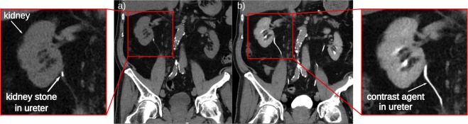
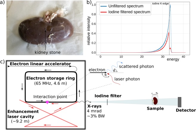
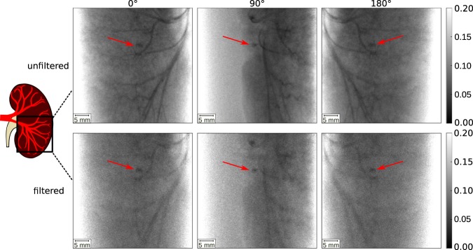
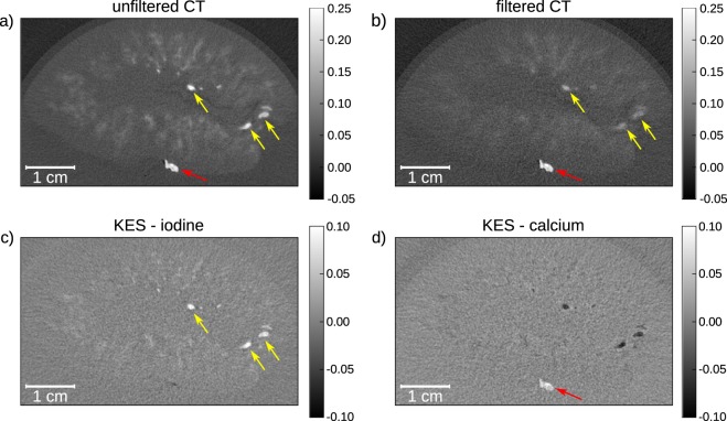
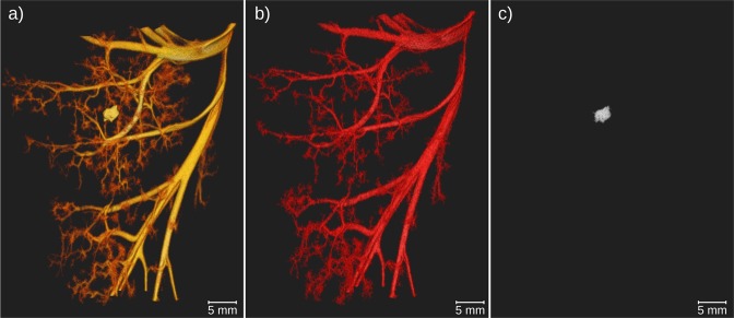
In clinical diagnosis, X-ray computed tomography (CT) is one of the most important imaging techniques. Yet, this method lacks the ability to differentiate similarly absorbing substances like commonly used iodine contrast agent and calcium which is typically seen in calcifications, kidney stones and bones. K-edge subtraction (KES) imaging can help distinguish these materials by subtracting two CT scans recorded at different X-ray energies. So far, this method mostly relies on monochromatic X-rays produced at large synchrotron facilities. Here, we present the first proof-of-principle experiment of a filter-based KES CT method performed at a compact synchrotron X-ray source based on inverse-Compton scattering, the Munich Compact Light Source (MuCLS). It is shown that iodine contrast agent and calcium can be clearly separated to provide CT volumes only showing one of the two materials. These results demonstrate that KES CT at a compact synchrotron source can become an important tool in pre-clinical research.
在临床诊断中,X 射线计算机断层扫描(CT)是最重要的成像技术之一。然而,这种方法缺乏区分类似吸收物质的能力,例如常用的碘造影剂和钙,这些物质通常存在于钙化、肾结石和骨骼中。K 边差减(KES)成像可以通过减去在不同 X 射线能量下记录的两个 CT 扫描来帮助区分这些材料。到目前为止,这种方法主要依赖于在大型同步加速器设施中产生的单色 X 射线。在这里,我们展示了在基于逆康普顿散射的紧凑型同步加速器 X 射线源(慕尼黑紧凑型光源,MuCLS)上进行的基于滤波器的 KES CT 方法的第一个原理验证实验。结果表明,可以清楚地分离碘造影剂和钙,从而提供仅显示两种材料之一的 CT 体积。这些结果表明,紧凑型同步加速器源的 KES CT 可以成为临床前研究中的重要工具。