Zeedan Gamil Sayed Gamil, Mahmoud Ayman Hamid, Abdalhamed Abeer Mostafa, El-Razik Khaled Abd El-Hamid Abd, Khafagi Manal Hamdy, Zeina Hala Abdoula Ahmed Abou
Department of Parasitology and Animals Diseases, National Research Centre, 33 Bohouth St., Dokki, Giza, P.O. Box 12622, Egypt.
Department of Biotechnology and Food Hygiene, Animal Health Institute, Dokki, Giza, Egypt.
Vet World. 2019 Jul;12(7):1093-1100. doi: 10.14202/vetworld.2019.1093-1100. Epub 2019 Jul 24.
Lumpy skin disease (LSD), is a highly infectious viral disease of cattle, caused by LSD virus (LSDV) which belongs to the genus of family . In the summer of 2017, skin lesions suggestive of LSD were observed in cattle at several governorates in Egypt. This study aimed to detect LSDV in cattle specimens using rapid serological and molecular diagnostic assays.
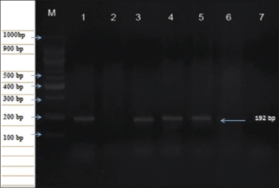
A total of 46 skin biopsies and uncoagulated blood samples were collected from cattle with LSD suggestive clinical signs, as well as 290 coagulated whole blood samples from cattle without skin lesion in different governorates in Egypt during the summer of 2017. Skin biopsies were used for virus isolation from the chorioallantoic membrane of 11-day-old specific pathogen-free embryonated chicken eggs (SPF-ECEs). LSDV was identified using conventional polymerase chain reaction (PCR), real-time PCR (RT-PCR), and fluorescent antibody technique (FAT) with specific hyperimmune serum against LSDV. Cattle sera were examined using indirect FAT (IFAT) and indirect enzyme-linked immunosorbent assay (ELISA).
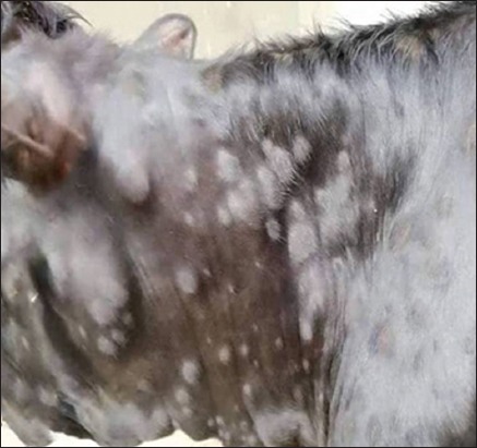
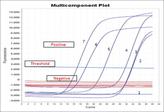
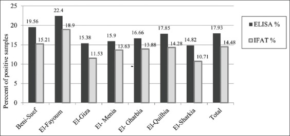
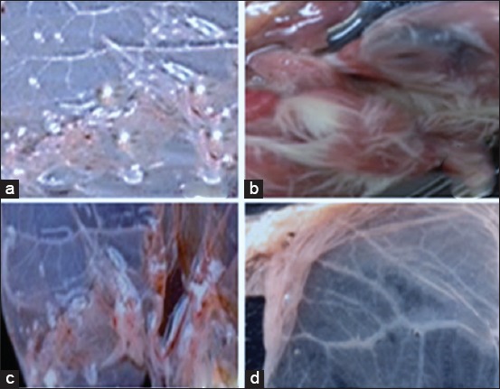
Skin nodules and sitfast lesions were significant clinical signs observed in all LSD suspect cattle. SPF-ECEs, from which positive isolations were made and it showed characteristic inflammatory and focal white pock lesions. The isolated viruses were identified as LSDV by FAT, conventional gel-based PCR, and RT-PCR. Among the skin biopsies and corresponding blood samples, LSDV-positive samples percentage were 39.13 and 36.95 by RT-PCR, followed 34.78 and 28.26 by conventional PCR and then 32.6 and 26.8 by FAT, respectively. The total positive percentage of LSDV antibody detected in cattle serum samples were 17.93 and 14.48 by indirect ELISA and IFAT.
LSDV was detected and identified in skin biopsies and corresponding blood samples of naturally infected cattle, more LSDV-positive samples were detected by RT-PCR, followed by conventional PCR and then FAT. The indirect ELISA detected more antibody-positive samples than the IFAT from cattle serum samples. The RT-PCR assay is simple, sensitive, rapid, and reliable for the detection of LSDV in blood and skin nodule biopsies of suspected cattle.
牛结节性皮肤病(LSD)是由牛结节性皮肤病病毒(LSDV)引起的一种牛的高度传染性病毒性疾病,LSDV属于 科的 属。2017年夏季,在埃及的几个省份的牛身上观察到疑似LSD的皮肤病变。本研究旨在使用快速血清学和分子诊断方法检测牛样本中的LSDV。
2017年夏季,从具有疑似LSD临床症状的牛身上采集了46份皮肤活检组织和未凝固血液样本,以及从埃及不同省份无皮肤病变的牛身上采集了290份凝固全血样本。皮肤活检组织用于从11日龄无特定病原体的鸡胚(SPF-ECEs)的绒毛尿囊膜中分离病毒。使用常规聚合酶链反应(PCR)、实时荧光定量PCR(RT-PCR)以及用抗LSDV的特异性超免疫血清进行荧光抗体技术(FAT)来鉴定LSDV。使用间接FAT(IFAT)和间接酶联免疫吸附测定(ELISA)检测牛血清。
在所有疑似LSD的牛中观察到皮肤结节和固着性病变是显著的临床症状。从中分离出阳性病毒的SPF-ECEs出现了特征性炎症和局灶性白色痘疱病变。通过FAT、常规凝胶PCR和RT-PCR将分离出的病毒鉴定为LSDV。在皮肤活检组织和相应血液样本中,RT-PCR检测到的LSDV阳性样本百分比分别为39.13%和36.95%,其次常规PCR分别为34.78%和28.26%,然后FAT分别为32.6%和26.8%。间接ELISA和IFAT检测到的牛血清样本中LSDV抗体总阳性百分比分别为17.93%和14.48%。
在自然感染牛的皮肤活检组织和相应血液样本中检测到并鉴定出LSDV,RT-PCR检测到的LSDV阳性样本更多,其次是常规PCR,然后是FAT。间接ELISA检测到的牛血清样本中抗体阳性样本比IFAT更多。RT-PCR检测方法对于检测疑似感染牛的血液和皮肤结节活检组织中的LSDV简单、灵敏、快速且可靠。