• 文献检索
  • 文档翻译
  • 深度研究
  • 学术资讯
  • Suppr Zotero 插件Zotero 插件
  • 邀请有礼
  • 套餐&价格
  • 历史记录
应用&插件
Suppr Zotero 插件Zotero 插件浏览器插件Mac 客户端Windows 客户端微信小程序
定价
高级版会员购买积分包购买API积分包
服务
文献检索文档翻译深度研究API 文档MCP 服务
关于我们
关于 Suppr公司介绍联系我们用户协议隐私条款
关注我们

Suppr 超能文献

核心技术专利:CN118964589B侵权必究
粤ICP备2023148730 号-1Suppr @ 2026

文献检索

告别复杂PubMed语法,用中文像聊天一样搜索,搜遍4000万医学文献。AI智能推荐,让科研检索更轻松。

立即免费搜索

文件翻译

保留排版,准确专业,支持PDF/Word/PPT等文件格式,支持 12+语言互译。

免费翻译文档

深度研究

AI帮你快速写综述,25分钟生成高质量综述,智能提取关键信息,辅助科研写作。

立即免费体验

一种新颖的深度学习架构在自动评估乳腺钼靶密度方面优于“现成的”迁移学习和基于特征的方法。

A novel deep learning architecture outperforming 'off‑the‑shelf' transfer learning and feature‑based methods in the automated assessment of mammographic breast density.

机构信息

Computational BioMedicine Laboratory (CBML), Institute of Computer Science (ICS), Foundation for Research and Technology‑Hellas (FORTH), 70013 Heraklion, Greece.

Department of Forensic Sciences and Laboratory of Toxicology, Medical School, University of Crete, 70013 Heraklion, Greece.

出版信息

Oncol Rep. 2019 Nov;42(5):2009-2015. doi: 10.3892/or.2019.7312. Epub 2019 Sep 12.

DOI:10.3892/or.2019.7312
PMID:31545461
原文链接:https://pmc.ncbi.nlm.nih.gov/articles/PMC6787954/
Abstract

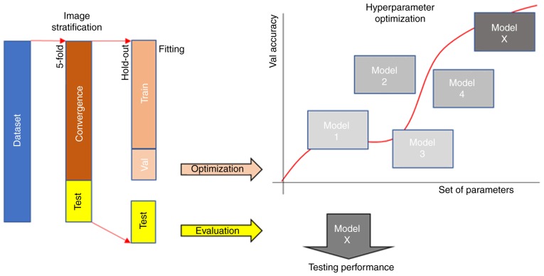
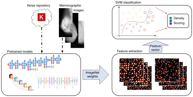
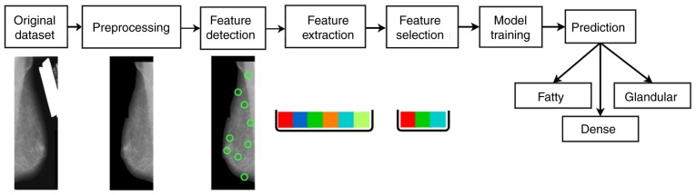
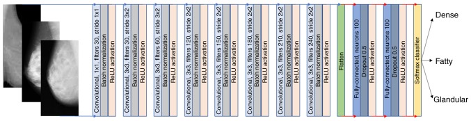
Potentially suspicious breast neoplasms could be masked by high tissue density, thus increasing the probability of a false‑negative diagnosis. Furthermore, differentiating breast tissue type enables patient pre‑screening stratification and risk assessment. In this study, we propose and evaluate advanced machine learning methodologies aiming at an objective and reliable method for breast density scoring from routine mammographic images. The proposed image analysis pipeline incorporates texture [Gabor filters and local binary pattern (LBP)] and gradient‑based features [histogram of oriented gradients (HOG) as well as speeded‑up robust features (SURF)]. Additionally, transfer learning approaches with ImageNet trained weights were also used for comparison, as well as a convolutional neural network (CNN). The proposed CNN model was fully trained on two open mammography datasets and was found to be the optimal performing methodology (AUC up to 87.3%). Thus, the findings of this study indicate that automated density scoring in mammograms can aid clinical diagnosis by introducing artificial intelligence‑powered decision‑support systems and contribute to the 'democratization' of healthcare by overcoming limitations, such as the geographic location of patients or the lack of expert radiologists.

摘要

高组织密度可能会掩盖潜在可疑的乳腺肿瘤,从而增加假阴性诊断的概率。此外,区分乳腺组织类型可以实现患者的预筛选分层和风险评估。在这项研究中,我们提出并评估了先进的机器学习方法,旨在为常规乳腺 X 光图像的乳腺密度评分提供一种客观可靠的方法。所提出的图像分析管道结合了纹理[Gabor 滤波器和局部二值模式(LBP)]和基于梯度的特征[方向梯度直方图(HOG)以及加速稳健特征(SURF)]。此外,还使用了带有 ImageNet 训练权重的迁移学习方法进行比较,以及卷积神经网络(CNN)。所提出的 CNN 模型在两个开放的乳腺 X 光数据集上进行了全面训练,被发现是表现最佳的方法(AUC 高达 87.3%)。因此,这项研究的结果表明,通过引入人工智能驱动的决策支持系统,对乳腺 X 光片进行自动密度评分可以辅助临床诊断,并通过克服患者地理位置或缺乏专家放射科医生等限制,为医疗保健的“民主化”做出贡献。

https://cdn.ncbi.nlm.nih.gov/pmc/blobs/4f79/6787954/da6189d24475/or-42-05-2009-g03.jpg
https://cdn.ncbi.nlm.nih.gov/pmc/blobs/4f79/6787954/75ac9cdd30de/or-42-05-2009-g00.jpg
https://cdn.ncbi.nlm.nih.gov/pmc/blobs/4f79/6787954/8ad207300ae8/or-42-05-2009-g01.jpg
https://cdn.ncbi.nlm.nih.gov/pmc/blobs/4f79/6787954/216234607933/or-42-05-2009-g02.jpg
https://cdn.ncbi.nlm.nih.gov/pmc/blobs/4f79/6787954/da6189d24475/or-42-05-2009-g03.jpg

相似文献

1
A novel deep learning architecture outperforming 'off‑the‑shelf' transfer learning and feature‑based methods in the automated assessment of mammographic breast density.一种新颖的深度学习架构在自动评估乳腺钼靶密度方面优于“现成的”迁移学习和基于特征的方法。
Oncol Rep. 2019 Nov;42(5):2009-2015. doi: 10.3892/or.2019.7312. Epub 2019 Sep 12.
2
Discriminating solitary cysts from soft tissue lesions in mammography using a pretrained deep convolutional neural network.使用预训练的深度卷积神经网络在乳腺钼靶摄影中区分孤立性囊肿与软组织病变。
Med Phys. 2017 Mar;44(3):1017-1027. doi: 10.1002/mp.12110.
3
Deep-Learning-Based Semantic Labeling for 2D Mammography and Comparison of Complexity for Machine Learning Tasks.基于深度学习的 2D 乳腺 X 光图像语义标注及机器学习任务复杂度比较。
J Digit Imaging. 2019 Aug;32(4):565-570. doi: 10.1007/s10278-019-00244-w.
4
Computer-aided assessment of breast density: comparison of supervised deep learning and feature-based statistical learning.计算机辅助乳腺密度评估:监督式深度学习与基于特征的统计学习方法的比较。
Phys Med Biol. 2018 Jan 9;63(2):025005. doi: 10.1088/1361-6560/aa9f87.
5
Large scale deep learning for computer aided detection of mammographic lesions.基于大规模深度学习的计算机辅助乳腺病变检测
Med Image Anal. 2017 Jan;35:303-312. doi: 10.1016/j.media.2016.07.007. Epub 2016 Aug 2.
6
Breast Cancer Classification in Automated Breast Ultrasound Using Multiview Convolutional Neural Network with Transfer Learning.基于多视图卷积神经网络和迁移学习的自动乳腺超声乳腺癌分类。
Ultrasound Med Biol. 2020 May;46(5):1119-1132. doi: 10.1016/j.ultrasmedbio.2020.01.001. Epub 2020 Feb 12.
7
Understanding Clinical Mammographic Breast Density Assessment: a Deep Learning Perspective.理解临床乳腺 X 线摄影乳房密度评估:深度学习视角。
J Digit Imaging. 2018 Aug;31(4):387-392. doi: 10.1007/s10278-017-0022-2.
8
Breast density prediction from low and standard dose mammograms using deep learning: effect of image resolution and model training approach on prediction quality.基于深度学习的低剂量和标准剂量乳腺钼靶图像的乳腺密度预测:图像分辨率和模型训练方法对预测质量的影响。
Biomed Phys Eng Express. 2024 May 15;10(4). doi: 10.1088/2057-1976/ad470b.
9
Comparison of a Deep Learning Risk Score and Standard Mammographic Density Score for Breast Cancer Risk Prediction.深度学习风险评分与标准乳腺密度评分在乳腺癌风险预测中的比较。
Radiology. 2020 Feb;294(2):265-272. doi: 10.1148/radiol.2019190872. Epub 2019 Dec 17.
10
Mass detection in digital breast tomosynthesis: Deep convolutional neural network with transfer learning from mammography.数字乳腺断层合成中的肿块检测:基于乳腺X线摄影迁移学习的深度卷积神经网络
Med Phys. 2016 Dec;43(12):6654. doi: 10.1118/1.4967345.

引用本文的文献

1
Investigating the value of radiomics stemming from DSC quantitative biomarkers in IDH mutation prediction in gliomas.研究源自DSC定量生物标志物的影像组学在胶质瘤IDH突变预测中的价值。
Front Neurol. 2023 Nov 16;14:1249452. doi: 10.3389/fneur.2023.1249452. eCollection 2023.
2
HER2 classification in breast cancer cells: A new explainable machine learning application for immunohistochemistry.乳腺癌细胞中的HER2分类:一种用于免疫组织化学的新型可解释机器学习应用。
Oncol Lett. 2022 Dec 14;25(2):44. doi: 10.3892/ol.2022.13630. eCollection 2023 Feb.
3
Artificial intelligence and convolution neural networks assessing mammographic images: a narrative literature review.

本文引用的文献

1
Mammographic Breast Density Assessment Using Deep Learning: Clinical Implementation.基于深度学习的乳腺 X 线摄影密度评估:临床应用。
Radiology. 2019 Jan;290(1):52-58. doi: 10.1148/radiol.2018180694. Epub 2018 Oct 16.
2
Mammographic density and breast cancer risk in breast screening assessment cases and women with a family history of breast cancer.乳腺筛查评估病例和有乳腺癌家族史的女性的乳腺密度与乳腺癌风险。
Eur J Cancer. 2018 Jan;88:48-56. doi: 10.1016/j.ejca.2017.10.022. Epub 2017 Nov 27.
3
A deep learning method for classifying mammographic breast density categories.
人工智能和卷积神经网络评估乳腺 X 光图像:叙事文献综述。
J Med Radiat Sci. 2020 Jun;67(2):134-142. doi: 10.1002/jmrs.385. Epub 2020 Mar 5.
一种用于对乳腺钼靶图像的乳房密度类别进行分类的深度学习方法。
Med Phys. 2018 Jan;45(1):314-321. doi: 10.1002/mp.12683. Epub 2017 Dec 22.
4
Unsupervised Deep Learning Applied to Breast Density Segmentation and Mammographic Risk Scoring.无监督深度学习在乳腺密度分割和乳腺钼靶风险评分中的应用。
IEEE Trans Med Imaging. 2016 May;35(5):1322-1331. doi: 10.1109/TMI.2016.2532122. Epub 2016 Feb 18.
5
The relationship of mammographic density and age: implications for breast cancer screening.乳腺密度与年龄的关系:对乳腺癌筛查的影响。
AJR Am J Roentgenol. 2012 Mar;198(3):W292-5. doi: 10.2214/AJR.10.6049.
6
EdgeFlow: a technique for boundary detection and image segmentation.边缘流:一种用于边界检测和图像分割的技术。
IEEE Trans Image Process. 2000;9(8):1375-88. doi: 10.1109/83.855433.
7
Breast cancer risk factors in relation to breast density (United States).与乳腺密度相关的乳腺癌风险因素(美国)
Cancer Causes Control. 2006 Dec;17(10):1281-90. doi: 10.1007/s10552-006-0071-1.
8
Mammographic features and breast cancer risk: effects with time, age, and menopause status.乳腺钼靶特征与乳腺癌风险:随时间、年龄及绝经状态的影响
J Natl Cancer Inst. 1995 Nov 1;87(21):1622-9. doi: 10.1093/jnci/87.21.1622.