Laudicella Riccardo, Albano Domenico, Annunziata Salvatore, Calabrò Diletta, Argiroffi Giovanni, Abenavoli Elisabetta, Linguanti Flavia, Albano Domenico, Vento Antonio, Bruno Antonio, Alongi Pierpaolo, Bauckneht Matteo
Department of Biomedical and Dental Sciences and of Morpho-Functional Imaging, Nuclear Medicine Unit, University of Messina, 98125 Messina, Italy.
Department of Nuclear Medicine, University of Brescia and Spedali Civili Brescia, 25123 Brescia, Italy.
Cancers (Basel). 2019 Sep 22;11(10):1412. doi: 10.3390/cancers11101412.
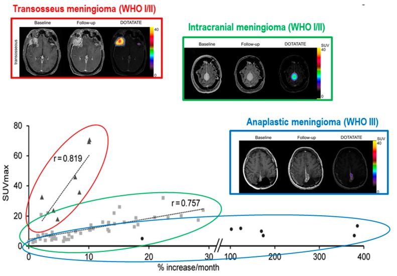
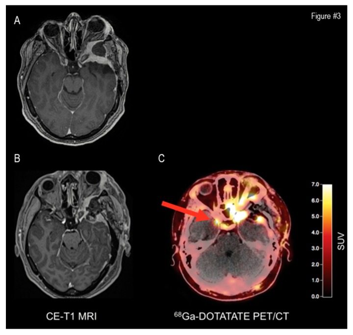
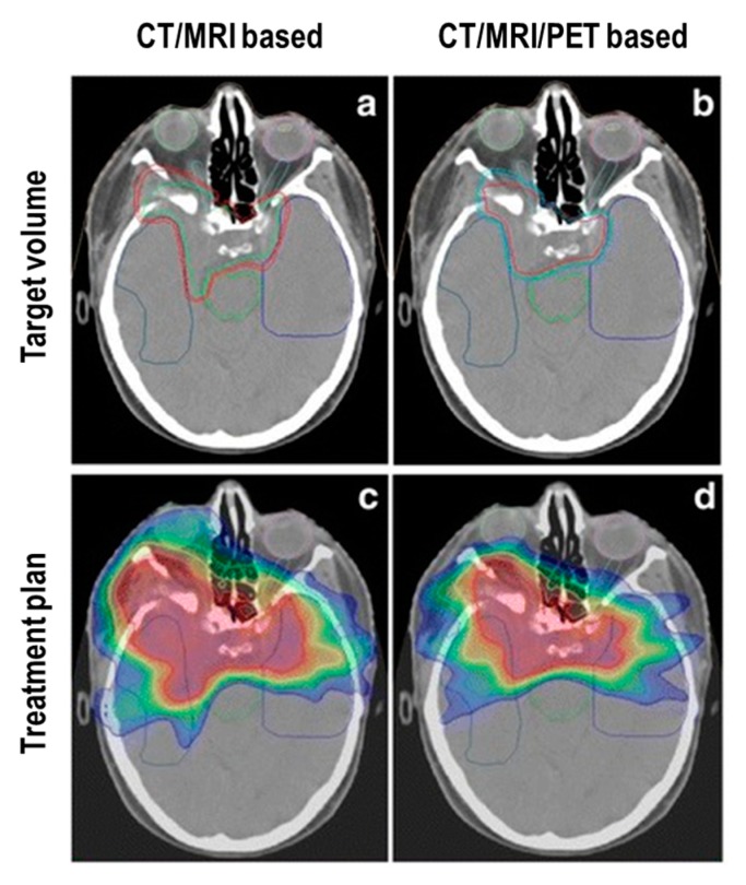
Meningiomas account for approximately 30% of all new diagnoses of intracranial masses. The 2016 World Health Organization's (WHO) classification currently represents the clinical standard for meningioma's grading and prognostic stratification. However, watchful waiting is frequently the chosen treatment option, although this means the absence of a certain histological diagnosis. Consequently, MRI (or less frequently CT) brain imaging currently represents the unique available tool to define diagnosis, grading, and treatment planning in many cases. Nonetheless, these neuroimaging modalities show some limitations, particularly in the evaluation of skull base lesions. The emerging evidence supporting the use of radiolabelled somatostatin receptor analogues (such as dota-peptides) to provide molecular imaging of meningiomas might at least partially overcome these limitations. Moreover, their potential therapeutic usage might enrich the current clinical offering for these patients. Starting from the strengths and weaknesses of structural and functional neuroimaging in meningiomas, in the present article we systematically reviewed the published studies regarding the use of radiolabelled dota-peptides in surgery and radiotherapy planning, in the restaging of treated patients, as well as in peptide-receptor radionuclide therapy of meningioma.
脑膜瘤约占所有新发颅内肿块诊断病例的30%。2016年世界卫生组织(WHO)的分类目前是脑膜瘤分级和预后分层的临床标准。然而,观察等待常常是首选的治疗方案,尽管这意味着缺乏明确的组织学诊断。因此,目前在许多情况下,头颅MRI(或较少使用的CT)成像仍是用于明确诊断、分级和制定治疗方案的唯一可用工具。尽管如此,这些神经影像学检查方法存在一些局限性,尤其是在评估颅底病变方面。支持使用放射性标记的生长抑素受体类似物(如多胺肽)对脑膜瘤进行分子成像的新证据可能至少部分克服这些局限性。此外,它们潜在的治疗用途可能会丰富目前为这些患者提供的临床治疗手段。本文从脑膜瘤结构和功能神经影像学的优缺点出发,系统回顾了已发表的关于放射性标记多胺肽在手术和放疗计划、治疗后患者再分期以及脑膜瘤肽受体放射性核素治疗中的应用研究。