Stem Genomics, IRMB, Univ Montpellier, INSERM, CHU Montpellier, Montpellier, France.
BioSystems and Micromechanics, IRG, Singapore-MIT Alliance for Research and Technology, Singapore, Singapore.
Sci Rep. 2019 Sep 24;9(1):13782. doi: 10.1038/s41598-019-50198-w.
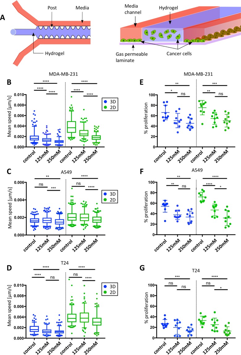
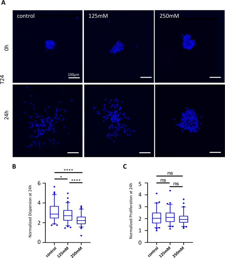
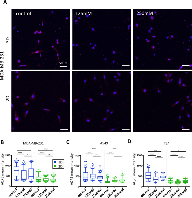
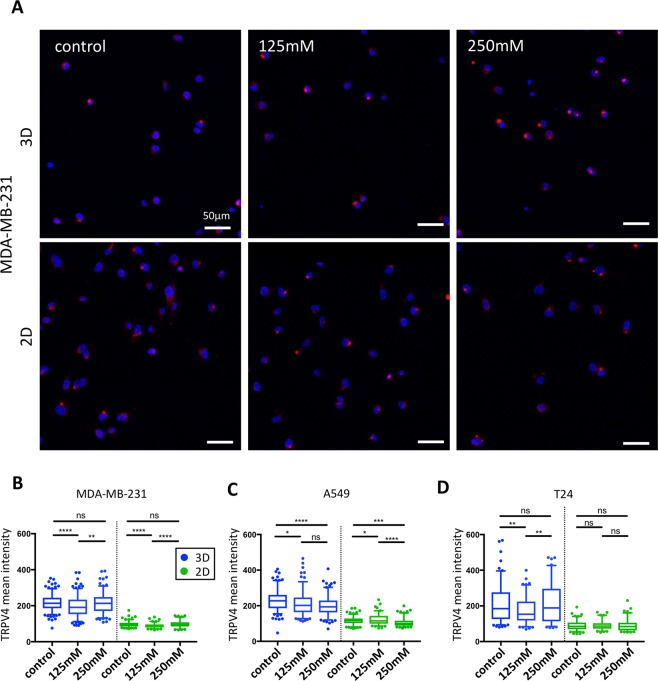
The maintenance of precise cell volume is critical for cell survival. Changes in extracellular osmolarity affect cell volume and may impact various cellular processes such as mitosis, mitochondrial functions, DNA repair as well as cell migration and proliferation. Much of what we know about the mechanisms of cell osmoregulation comes from in vitro two-dimensional (2D) assays that are less physiologically relevant than three-dimensional (3D) in vitro or in vivo settings. Here, we developed a microfluidic model to study the impact of hyper-osmotic stress on the migration, proliferation and ion channel/transporter expression changes of three metastatic cell lines (MDA-MB-231, A549, T24) in 2D versus 3D environments. We observed a global decrease in cell migration and proliferation upon hyper-osmotic stress treatment, with similar responses between 2D and 3D conditions. Specific ion channels/aquaporins are over-expressed in metastatic cells and play a central role during osmo-regulation. Therefore, the effects of hyper-osmotic stress on two transporters, aquaporin 5 (AQP5) and the transient receptor potential cation channel (TRPV4), was investigated. While hyper-osmotic stress had no major impact on the transporters of cells cultured in 2D, cells embedded in collagen gel (3D) decreased their AQP5 expression and exhibited a reduction in intra-cellular translocation of TRPV4. Furthermore, cell dispersion from T24 aggregates embedded in 3D collagen gel decreased with higher levels of hyper-osmotic stress. In conclusion, this study provides evidence on the impact of hyper-osmotic stress on various aspects of metastatic cell progression and highlights the importance of having a 3D cell culture platform in investigating molecular players involved in cancer cell migration.
精确细胞体积的维持对于细胞存活至关重要。细胞外渗透压的变化会影响细胞体积,并可能影响各种细胞过程,如有丝分裂、线粒体功能、DNA 修复以及细胞迁移和增殖。我们对细胞渗透调节机制的了解主要来自于体外二维(2D)测定,这些测定与三维(3D)体外或体内环境相比,生理相关性较低。在这里,我们开发了一种微流控模型,以研究高渗应激对三种转移细胞系(MDA-MB-231、A549、T24)在 2D 与 3D 环境中迁移、增殖和离子通道/转运体表达变化的影响。我们观察到高渗应激处理后细胞迁移和增殖的整体下降,2D 和 3D 条件下的反应相似。特定的离子通道/水通道在转移性细胞中过表达,并在渗透调节中发挥核心作用。因此,研究了高渗应激对两种转运蛋白,水通道蛋白 5(AQP5)和瞬时受体电位阳离子通道(TRPV4)的影响。虽然高渗应激对 2D 培养细胞的转运体没有重大影响,但嵌入胶原凝胶(3D)的细胞减少了 AQP5 的表达,并表现出 TRPV4 细胞内易位减少。此外,随着高渗应激水平的升高,嵌入 3D 胶原凝胶中的 T24 聚集体的细胞分散减少。总之,这项研究提供了高渗应激对转移细胞进展各个方面的影响的证据,并强调了在研究参与癌细胞迁移的分子参与者时使用 3D 细胞培养平台的重要性。