Saad Amin F, Chaudhari Ruchir, Wintermark Max
Department of Radiology, Baylor University Medical Center, Dallas, TX, United States.
Department of Radiology, Stanford University, Stanford, CA, United States.
Front Neurol. 2019 Sep 4;10:964. doi: 10.3389/fneur.2019.00964. eCollection 2019.
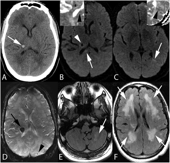
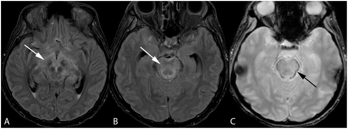
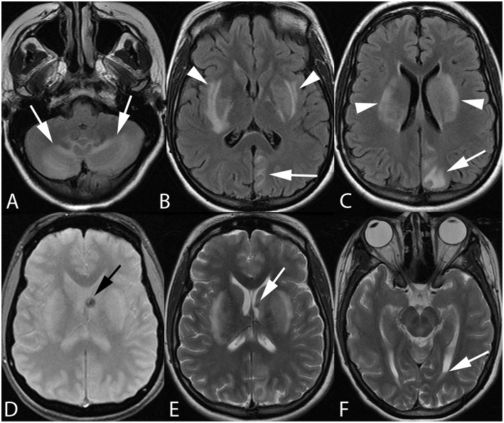
Posterior reversible encephalopathy syndrome (PRES) is a condition clinically characterized by headache, altered mental status, seizures, and visual loss and may be associated with systemic hypertension, preeclampsia/eclampsia, chemotherapy, immunosuppressive therapies in the setting of organ transplantation, and uremic encephalopathy. While brain imaging in patients with PRES typically reveals symmetric vasogenic edema within the parietal and occipital lobes, PRES may present with atypical imaging findings such as central brainstem and deep gray involvement without subcortical edema, and even spinal cord involvement. Additionally, PRES may be complicated in some cases by the presence of cytotoxic edema and hemorrhage. This review will serve to summarize the pathophysiologic theories and controversies underlying PRES, imaging features encountered in atypical and complicated PRES, and the implications these findings may have on patient prognosis.
后部可逆性脑病综合征(PRES)是一种临床特征为头痛、精神状态改变、癫痫发作和视力丧失的病症,可能与系统性高血压、先兆子痫/子痫、化疗、器官移植背景下的免疫抑制治疗以及尿毒症性脑病有关。虽然PRES患者的脑部成像通常显示顶叶和枕叶内对称的血管源性水肿,但PRES可能表现出非典型的成像结果,如脑干中央和深部灰质受累而无皮质下水肿,甚至脊髓受累。此外,在某些情况下,PRES可能并发细胞毒性水肿和出血。本综述旨在总结PRES潜在的病理生理理论和争议、非典型和复杂PRES中遇到的成像特征,以及这些发现可能对患者预后产生的影响。