Yasokawa Naoya, Shirai Ryo, Tanaka Hitomi, Kurose Koji, Oga Toru, Oka Mikio
Department of Respiratory Medicine, Kawasaki Medical School, Japan.
Department of Immuno-Oncology, Kawasaki Medical School, Japan.
Intern Med. 2020 Jan 15;59(2):257-260. doi: 10.2169/internalmedicine.3031-19. Epub 2019 Sep 26.
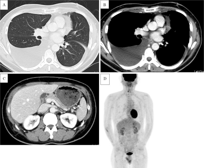
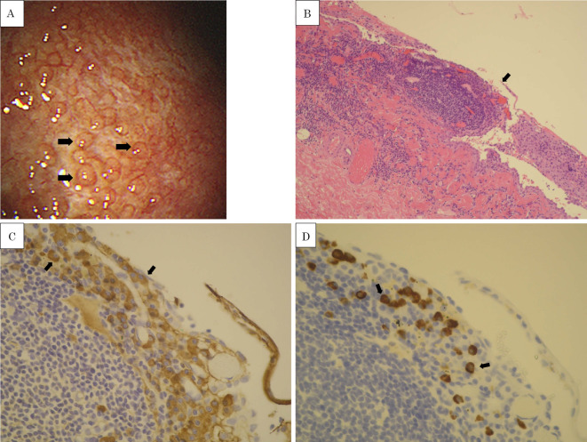
A 46-year-old Japanese man was admitted to our hospital with a 1-year history of dyspnea and persistent right-dominant bilateral pleural effusions. Chest and abdominal computed tomography (CT) revealed no notable findings apart from the bilateral pleural effusions. 2-deoxy-2-[F]-fluoro-D-glucose (FDG) positron emission tomography-CT showed no accumulation of FDG in the thorax and abdomen. Thoracoscopy revealed numerous small (approximately 2-3 mm in size), blister-like nodules on the left parietal pleura extending from the lower third of the chest wall to the diaphragm. A pathological examination revealed lymphocyte and plasma cell infiltrates with increasing numbers of IgG4-positive plasma cells in the fibrotic pleura, indicating IgG4-related pleuritis.
一名46岁的日本男性因呼吸困难病史1年且持续性右侧优势双侧胸腔积液入住我院。胸部和腹部计算机断层扫描(CT)除双侧胸腔积液外未发现明显异常。2-脱氧-2-[F]-氟-D-葡萄糖(FDG)正电子发射断层扫描-CT显示胸部和腹部无FDG聚集。胸腔镜检查发现左壁层胸膜有许多小的(大小约2-3毫米)水泡样结节,从胸壁下三分之一延伸至膈肌。病理检查显示纤维化胸膜中有淋巴细胞和浆细胞浸润,IgG4阳性浆细胞数量增加,提示IgG4相关性胸膜炎。