Lee Hyunjin, Park Jun-Beom
Department of Periodontics, College of Medicine, The Catholic University of Korea, Seoul, Republic of Korea.
Eur J Dent. 2019 May;13(2):131-136. doi: 10.1055/s-0039-1694904. Epub 2019 Oct 1.
Dimethyl sulfoxide (DMSO) plays various functions, including cellular functions such as cellular growth. The aim of this study was to evaluate the effects of DMSO on the proliferation and osteogenic differentiation of human gingiva-derived stem cells.
Stem cells derived from gingiva were cultured in the presence of DMSO at concentrations ranging from 0.01 to 10%.
We performed a one-way analysis of variance (ANOVA) with post hoc test to determine the differences between the groups using a commercially available program and the level of significance was 0.05.
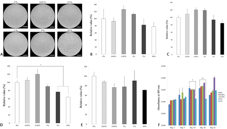
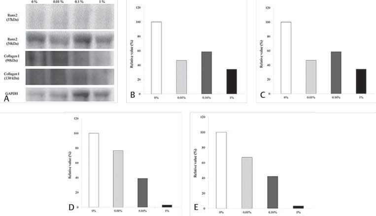
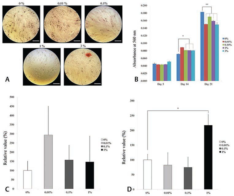
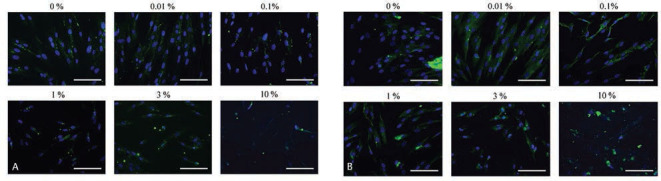
The cells in the control group showed normal fibroblast morphology. The cells treated with 0.01%, 0.01%, 0.1%, and 1% DMSO were morphologically similar to those from the control group on each day. Statistically significant decreases in cell counting kit-8 (CCK-8) values were seen in the 3% and 10% DMSO groups ( 0.05). A statistically significant decrease in alkaline phosphatase activity was seen in the 3% DMSO group. ( 0.05). The application of DMSO produced a decrease in alizarin red S staining. The expression of Runx2 and collagen I by immunofluorescence decreased as the dose of lovastatin increased.
The effects of DMSO on the viability of osteogenic differentiation among stem cells derived from human gingiva were evaluated. Applying DMSO produced decreased cell viability and decreased osteogenic differentiation in this experimental setting. This should be considered when designing and interpreting the data, and a DMSO-free method may be considered for bone regeneration applications.
二甲基亚砜(DMSO)具有多种功能,包括细胞生长等细胞功能。本研究的目的是评估DMSO对人牙龈来源干细胞增殖和成骨分化的影响。
将牙龈来源的干细胞在浓度范围为0.01%至10%的DMSO存在下进行培养。
我们使用市售程序进行单因素方差分析(ANOVA)并进行事后检验,以确定组间差异,显著性水平为0.05。
对照组细胞呈现正常成纤维细胞形态。在每天,用0.01%、0.1%和1% DMSO处理的细胞在形态上与对照组细胞相似。在3%和10% DMSO组中,细胞计数试剂盒-8(CCK-8)值出现统计学显著下降(P<0.05)。在3% DMSO组中,碱性磷酸酶活性出现统计学显著下降(P<0.05)。DMSO的应用导致茜素红S染色减少。随着洛伐他汀剂量增加,免疫荧光检测显示Runx2和I型胶原蛋白的表达下降。
评估了DMSO对人牙龈来源干细胞成骨分化活力的影响。在本实验环境中,应用DMSO导致细胞活力降低和成骨分化减少。在设计和解释数据时应考虑这一点,对于骨再生应用,可考虑采用无DMSO的方法。