Department of Ophthalmology, University of Missouri School of Medicine, Columbia, Missouri, USA
Department of Ophthalmology, University of Missouri School of Medicine, Columbia, Missouri, USA.
Br J Ophthalmol. 2020 Jul;104(7):999-1004. doi: 10.1136/bjophthalmol-2019-314466. Epub 2019 Oct 4.
BACKGROUND/AIMS: Meibomian gland dysfunction (MGD) is the most common form of evaporative dry eye disease, but its pathogenesis is poorly understood. This study examined the histopathological features of meibomian gland (MG) tissue from cadaver donors to identify potential pathogenic processes that underlie MGD in humans.
Histological analyses was performed on the MGs in the tarsal plates dissected from four cadaver donors, two young and two old adults, including a 36-year-old female (36F) and three males aged 30, 63 and 64 years (30M, 63M and 64M).
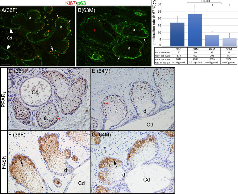
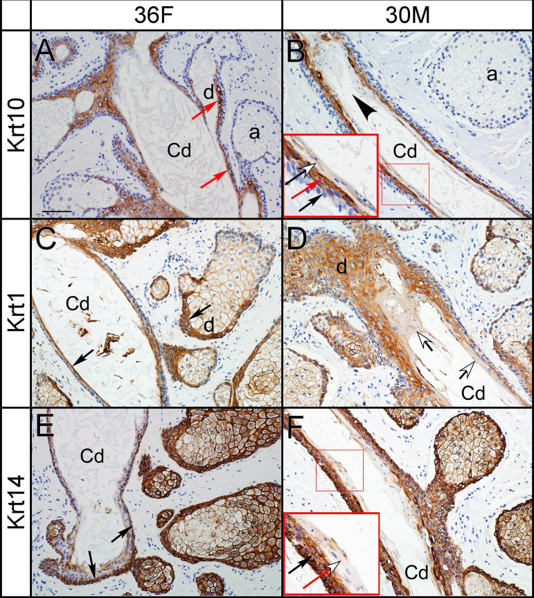
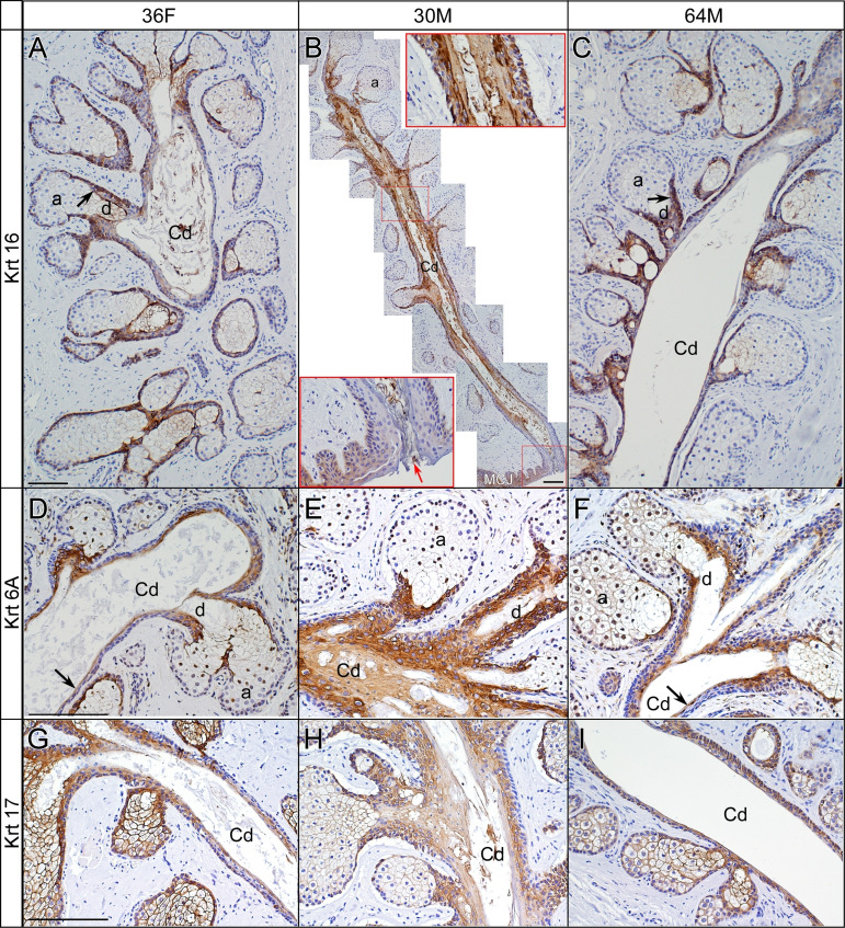
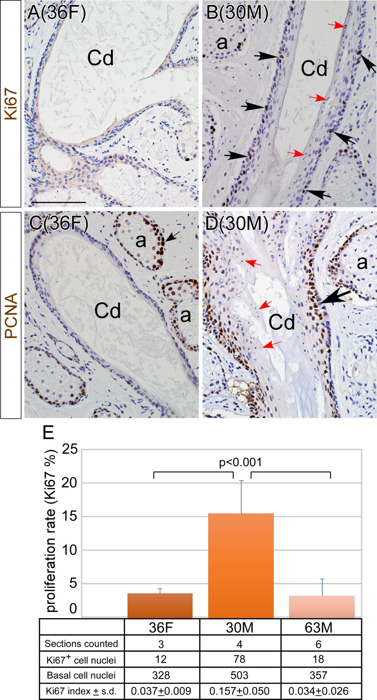
The MGs of 36F displayed normal anatomy and structure, whereas the MGs of 30M showed severe ductal obstruction with mild distortion. The obstruction was caused by increased cytokeratin levels in association with hyperproliferation, but not hyperkeratinisation. In two older males, moderate to severe MG atrophy was noted. Cell proliferation was significantly reduced in the MG acini of the two older donors as measured by Ki67 labelling index (6.0%±3.4% and 7.9%±2.8% in 63M and 64M, respectively) when compared with that of the two younger donors (23.2%±5.5% and 16.9%±4.8% in 30M and 36F, respectively) (p<0.001). The expression patterns of meibocyte differentiation biomarkers were similar in the older and younger donors.
Our histopathological study, based on a small sample size, suggests potentially distinct pathogenic mechanisms in MGD. In the young male adult, hyperproliferation and aberrant differentiation of the central ductal epithelia may lead to the obstruction by overproduced cytokeratins. In contrast, in older adults, decreased cell proliferation in acinar basal epithelia could be a contributing factor leading to MG glandular atrophy.
背景/目的:睑板腺功能障碍(MGD)是最常见的蒸发性干眼症形式,但发病机制尚不清楚。本研究通过检查尸检供体的睑板腺(MG)组织的组织病理学特征,确定了人类 MGD 潜在的发病机制。
对从四名尸检供体(两名年轻和两名老年成人)的睑板中分离出的 MG 进行组织学分析,两名年轻成人包括一名 36 岁女性(36F)和一名 30 岁男性(30M),两名老年男性分别为 63 岁和 64 岁(63M 和 64M)。
36F 的 MG 显示出正常的解剖结构和形态,而 30M 的 MG 则表现出严重的导管阻塞,伴有轻微的变形。阻塞是由于细胞角蛋白水平增加伴细胞过度增生引起的,但没有过度角化。在两名老年男性中,MG 明显萎缩。Ki67 标记指数显示,两名老年供体的 MG 腺泡细胞增殖显著减少(63M 和 64M 分别为 6.0%±3.4%和 7.9%±2.8%),与两名年轻供体(30M 和 36F 分别为 23.2%±5.5%和 16.9%±4.8%)相比(p<0.001)。在年轻和老年供体中,MEIBO 细胞分化标志物的表达模式相似。
我们的组织病理学研究表明,在 MGD 中可能存在不同的发病机制。在年轻男性中,中央导管上皮的过度增生和异常分化可能导致过度产生的细胞角蛋白阻塞。相比之下,在老年男性中,腺泡基底上皮细胞增殖减少可能是导致 MG 腺萎缩的一个因素。