Wang Qianqian, Zang Jingyu, Huang Xu, Lu Hongli, Xu Wenxie, Chen Jie
Department of Pediatric Surgery, Xinhua Hospital Affiliated to Shanghai Jiaotong University School of Medicine, Shanghai, China.
Department of Pediatric Surgery, Jiaxing Maternity and Child Health Care Hospital, Jiaxing, China.
J Neurogastroenterol Motil. 2019 Oct 30;25(4):589-601. doi: 10.5056/jnm19136.
BACKGROUND/AIMS: Interstitial cells play important roles in gastrointestinal (GI) neuro-smooth muscle transmission. The underlying mechanisms of colonic dysmotility have not been well illustrated. We established a partial colon obstruction (PCO) mouse model to investigate the changes of interstitial cells and the correlation with colonic motility.
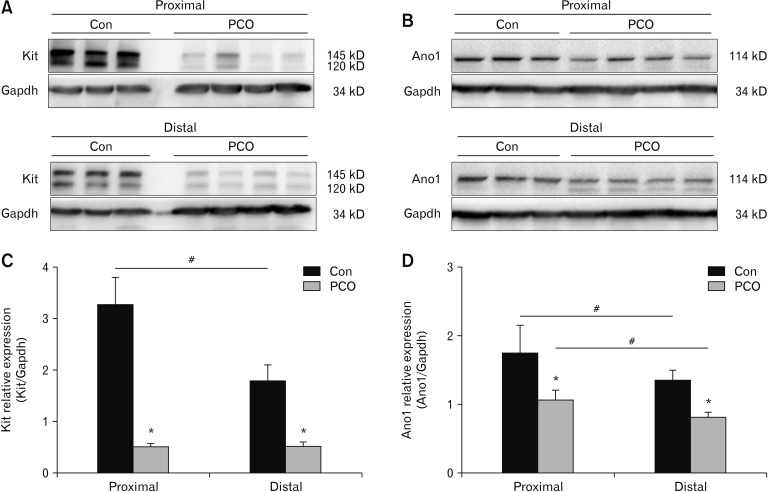
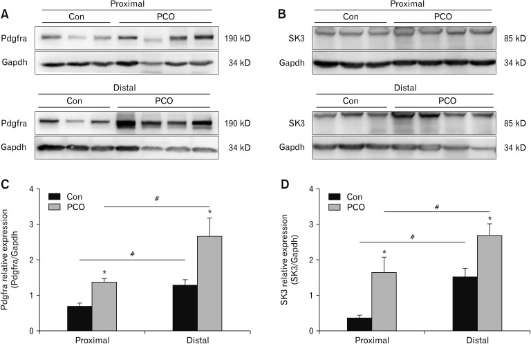
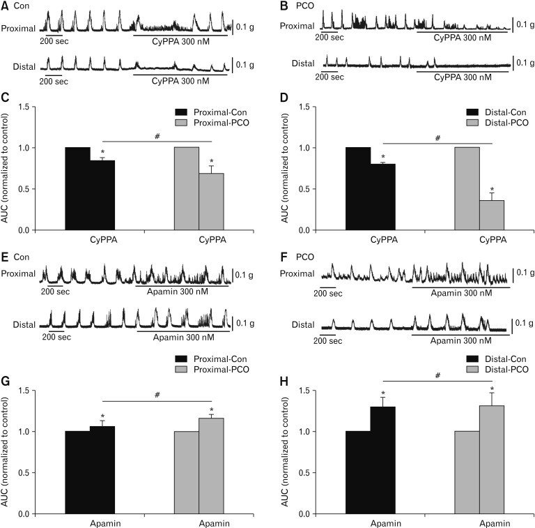
Western blot technique was employed to observe the protein expressions of Kit, platelet-derived growth factor receptor-α (Pdgfra), Ca-activated Cl (Ano1) channels, and small conductance Ca- activated K (SK) channels. Colonic migrating motor complexes (CMMCs) and isometric force measurements were employed in control mice and PCO mice.
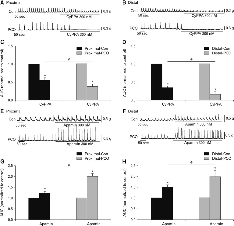
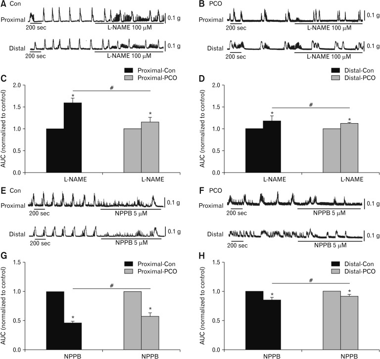
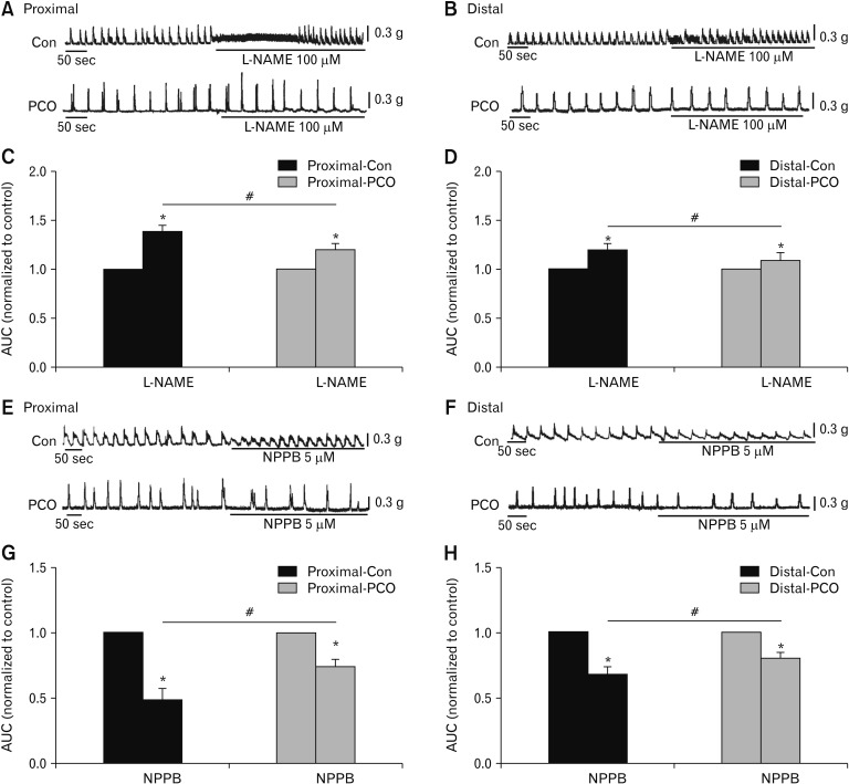
PCO mice showed distended abdomen and feces excretion was significantly reduced. Anatomically, the colon above the obstructive silicone ring was obviously dilated. Kit and Ano1 proteins in the colonic smooth muscle layer of the PCO colons were significantly decreased, while the expression of Pdgfra and SK3 proteins were significantly increased. The effects of a nitric oxide synthase inhibitor (L-NAME) and an Ano1 channel inhibitor (NPPB) on CMMC and colonic spontaneous contractions were decreased in the proximal and distal colons of PCO mice. The SK agonist, CyPPA and antagonist, apamin in PCO mice showed more effect to the CMMCs and colonic smooth muscle contractions.
Colonic transit disorder may be due to the downregulation of the Kit and Ano1 channels and the upregulation of SK3 channels in platelet-derived growth factor receptor-α positive (PDGFRα) cells. The imbalance between interstitial cells of Cajal-Ano1 and PDGFRαSK3 distribution might be a potential reason for the colonic dysmotility.
背景/目的:间质细胞在胃肠神经-平滑肌传递中起重要作用。结肠动力障碍的潜在机制尚未完全阐明。我们建立了部分结肠梗阻(PCO)小鼠模型,以研究间质细胞的变化及其与结肠动力的相关性。
采用蛋白质免疫印迹技术观察干细胞因子受体(Kit)、血小板衍生生长因子受体-α(Pdgfra)、钙激活氯离子通道(Ano1)和小电导钙激活钾通道(SK)的蛋白表达。对对照小鼠和PCO小鼠进行结肠移行性运动复合波(CMMC)和等长收缩力测量。
PCO小鼠出现腹部膨胀,粪便排泄显著减少。解剖学上,梗阻硅胶环上方的结肠明显扩张。PCO结肠的结肠平滑肌层中Kit和Ano1蛋白显著减少,而Pdgfra和SK3蛋白的表达显著增加。一氧化氮合酶抑制剂(L-NAME)和Ano1通道抑制剂(NPPB)对PCO小鼠近端和远端结肠CMMC和结肠自发收缩的作用减弱。SK激动剂CyPPA和拮抗剂蜂毒明肽对PCO小鼠的CMMC和结肠平滑肌收缩有更大影响。
结肠转运障碍可能是由于血小板衍生生长因子受体-α阳性(PDGFRα)细胞中Kit和Ano1通道下调以及SK3通道上调所致。 Cajal间质细胞-Ano1和PDGFRα-SK3分布失衡可能是结肠动力障碍的潜在原因。