Michael Smith Genome Sciences Centre, British Columbia Cancer Agency, 675W 10th Ave, Vancouver, BC, V5Z 1L3, Canada.
Department of Medical Genetics, University of British Columbia, C201 - 4500 Oak Street, Vancouver, BC, V6H 3N1, Canada.
Nat Commun. 2019 Oct 7;10(1):4553. doi: 10.1038/s41467-019-12444-7.
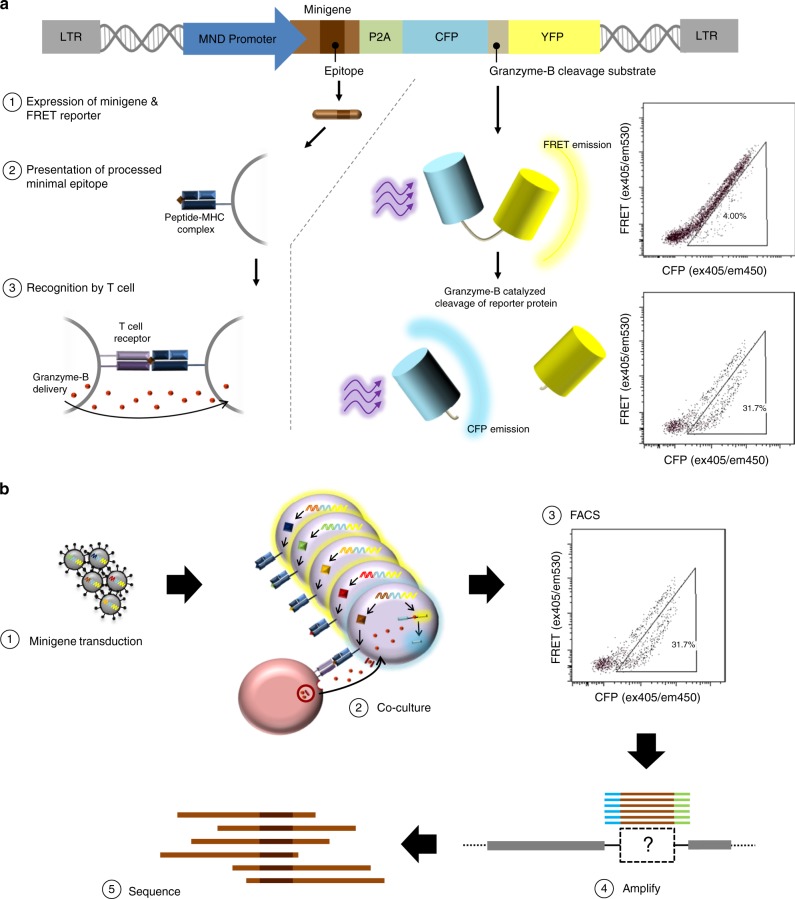
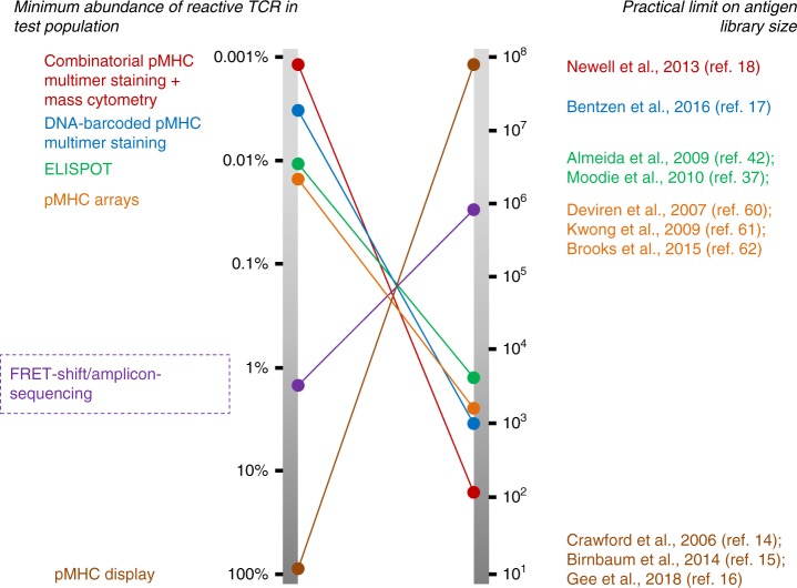
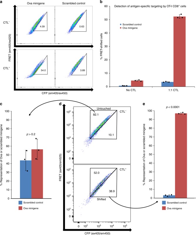
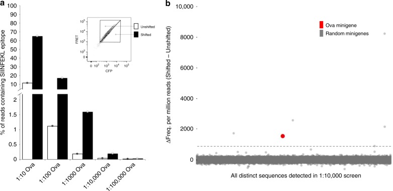
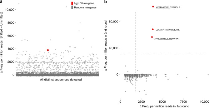
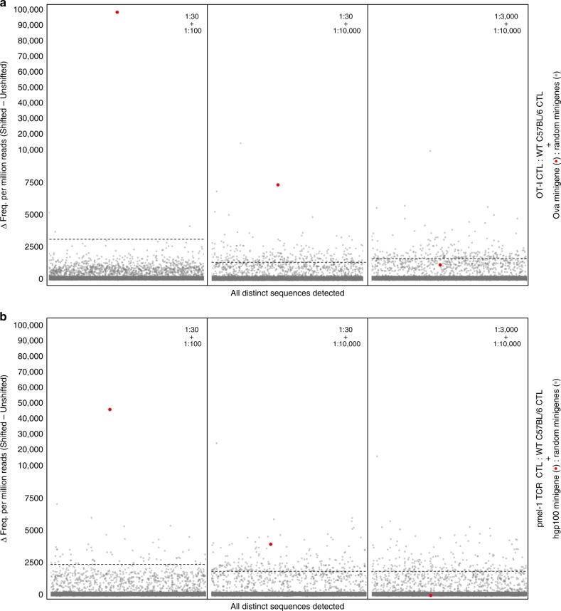
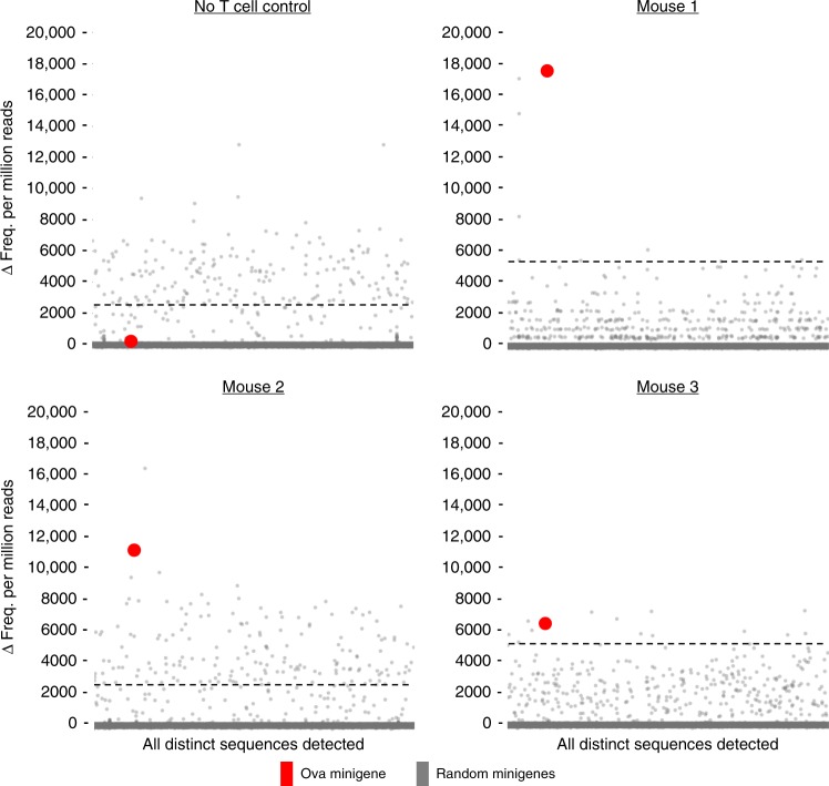
Cytotoxic CD8 T cells recognize and eliminate infected or malignant cells that present peptide epitopes derived from intracellularly processed antigens on their surface. However, comprehensive profiling of specific major histocompatibility complex (MHC)-bound peptide epitopes that are naturally processed and capable of eliciting a functional T cell response has been challenging. Here, we report a method for deep and unbiased T cell epitope profiling, using in vitro co-culture of CD8 T cells together with target cells transduced with high-complexity, epitope-encoding minigene libraries. Target cells that are subject to cytotoxic attack from T cells in co-culture are isolated prior to apoptosis by fluorescence-activated cell sorting, and characterized by sequencing the encoded minigenes. We then validate this highly parallelized method using known murine T cell receptor/peptide-MHC pairs and diverse minigene-encoded epitope libraries. Our data thus suggest that this epitope profiling method allows unambiguous and sensitive identification of naturally processed and MHC-presented peptide epitopes.
细胞毒性 CD8 T 细胞识别并消除表面呈现源自细胞内处理的抗原的肽表位的感染或恶性细胞。然而,对能够引发功能性 T 细胞反应的特定主要组织相容性复合体 (MHC) 结合肽表位进行全面分析一直具有挑战性。在这里,我们报告了一种使用体外共培养 CD8 T 细胞与转导高复杂度、表位编码的小基因文库的靶细胞进行深度和无偏的 T 细胞表位分析的方法。在共培养的 T 细胞对靶细胞进行细胞毒性攻击之前,通过荧光激活细胞分选将处于凋亡前的靶细胞分离,并通过测序编码的小基因进行表征。然后,我们使用已知的小鼠 T 细胞受体/肽-MHC 对和多样化的小基因编码的表位文库验证了这种高度并行的方法。因此,我们的数据表明,这种表位分析方法允许明确和敏感地识别天然加工和 MHC 呈递的肽表位。