Laboratory of Neurobiology and Stem Cells NeuroCellT, Department of Cellular Biology, Faculty of Biological Sciences, University of Concepcion, Concepcion, Chile.
Center for Advanced Microscopy CMA BIO BIO, University of Concepcion, Concepcion, Chile.
Sci Rep. 2019 Oct 8;9(1):14422. doi: 10.1038/s41598-019-50772-2.
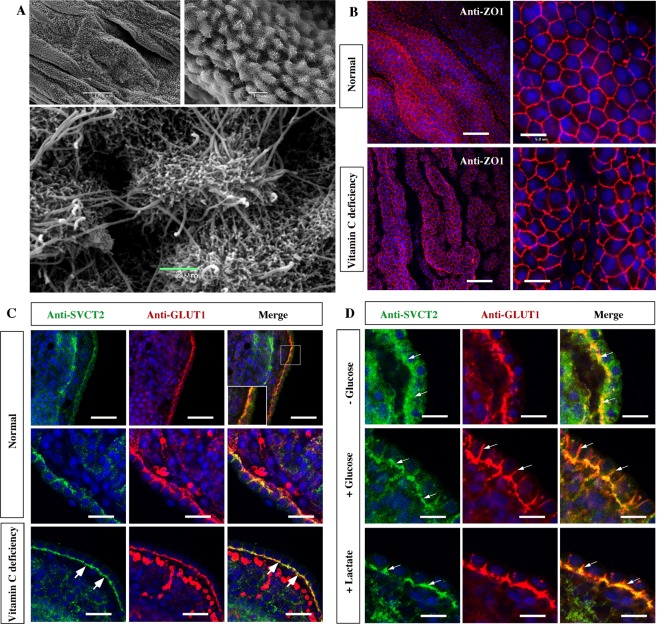
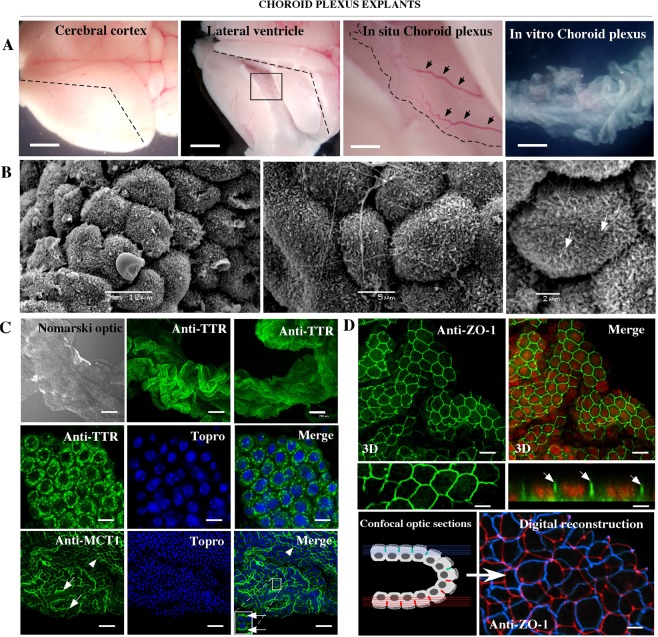
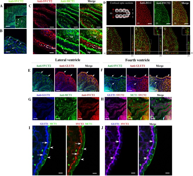
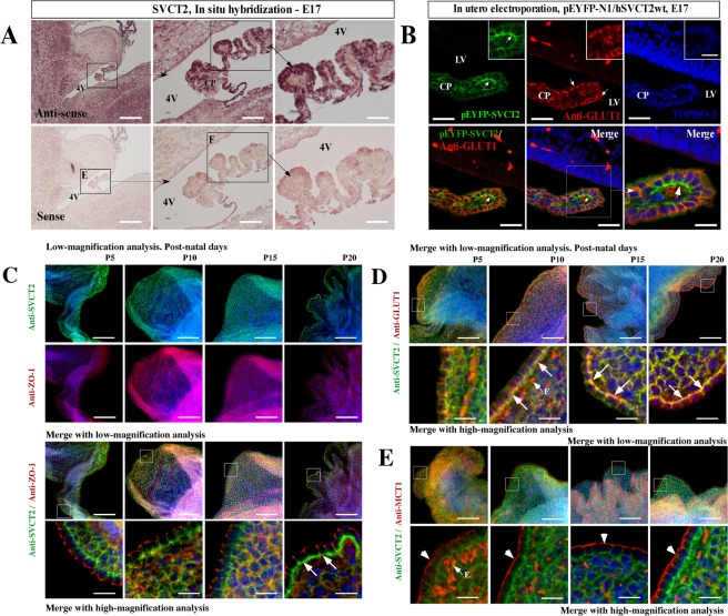
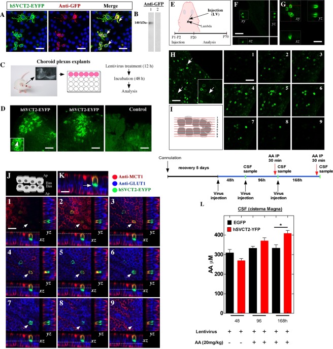
Vitamin C is incorporated into the cerebrospinal fluid (CSF) through choroid plexus cells. While the transfer of vitamin C from the blood to the brain has been studied functionally, the vitamin C transporter, SVCT2, has not been detected in the basolateral membrane of choroid plexus cells. Furthermore, it is unknown how its expression is induced in the developing brain and modulated in scurvy conditions. We concluded that SVCT2 is intensely expressed in the second half of embryonic brain development and postnatal stages. In postnatal and adult brain, SVCT2 is highly expressed in all choroidal plexus epithelial cells, shown by colocalization with GLUT1 in the basolateral membranes and without MCT1 colocalization, which is expressed in the apical membrane. We confirmed that choroid plexus explant cells (in vitro) form a sealed epithelial structure, which polarized basolaterally, endogenous or overexpressed SVCT2. These results are reproduced in vivo by injecting hSVCT2wt-EYFP lentivirus into the CSF. Overexpressed SVCT2 incorporates AA (intraperitoneally injected) from the blood to the CSF. Finally, we observed in Guinea pig brain under scorbutic condition, that normal distribution of SVCT2 in choroid plexus may be regulated by peripheral concentrations of vitamin C. Additionally, we observed that SVCT2 polarization also depends on the metabolic stage of the choroid plexus cells.
维生素 C 通过脉络丛细胞整合到脑脊液(CSF)中。虽然已经从功能上研究了维生素 C 从血液向大脑的转移,但尚未在脉络丛细胞的基底外侧膜上检测到维生素 C 转运蛋白 SVCT2。此外,其在发育中的大脑中的表达如何诱导以及在坏血病条件下如何调节尚不清楚。我们得出结论,SVCT2 在胚胎脑发育的后半期和出生后阶段强烈表达。在出生后和成年期的大脑中,SVCT2 在所有脉络丛上皮细胞中高度表达,通过与基底外侧膜中的 GLUT1 共定位来证明,而没有 MCT1 共定位,MCT1 在顶膜中表达。我们证实脉络丛外植体细胞(体外)形成密封的上皮结构,该结构具有基底外侧的极性,内源性或过表达的 SVCT2。通过将 hSVCT2wt-EYFP 慢病毒注入 CSF 中,在体内重现了这些结果。过表达的 SVCT2 将 AA(腹腔内注射)从血液转移到 CSF 中。最后,我们在坏血病状态下的豚鼠大脑中观察到,脉络丛中 SVCT2 的正常分布可能受外周维生素 C 浓度的调节。此外,我们还观察到 SVCT2 的极化也取决于脉络丛细胞的代谢阶段。