• 文献检索
  • 文档翻译
  • 深度研究
  • 学术资讯
  • Suppr Zotero 插件Zotero 插件
  • 邀请有礼
  • 套餐&价格
  • 历史记录
应用&插件
Suppr Zotero 插件Zotero 插件浏览器插件Mac 客户端Windows 客户端微信小程序
定价
高级版会员购买积分包购买API积分包
服务
文献检索文档翻译深度研究API 文档MCP 服务
关于我们
关于 Suppr公司介绍联系我们用户协议隐私条款
关注我们

Suppr 超能文献

核心技术专利:CN118964589B侵权必究
粤ICP备2023148730 号-1Suppr @ 2026

文献检索

告别复杂PubMed语法,用中文像聊天一样搜索,搜遍4000万医学文献。AI智能推荐,让科研检索更轻松。

立即免费搜索

文件翻译

保留排版,准确专业,支持PDF/Word/PPT等文件格式,支持 12+语言互译。

免费翻译文档

深度研究

AI帮你快速写综述,25分钟生成高质量综述,智能提取关键信息,辅助科研写作。

立即免费体验

使用心脏特异性柔性 3D 打印丙烯酸支架结合非刚性配准,提高离体和在体心血管磁共振图像的配准精度。

Improved co-registration of ex-vivo and in-vivo cardiovascular magnetic resonance images using heart-specific flexible 3D printed acrylic scaffold combined with non-rigid registration.

机构信息

School of Biomedical Engineering and Imaging Sciences, King's College, London, Fourth Floor Lambeth Wing, St Thomas' Hospital, London, SE1 7EH, UK.

Siemens Healthcare Limited, Frimley, UK.

出版信息

J Cardiovasc Magn Reson. 2019 Oct 10;21(1):62. doi: 10.1186/s12968-019-0574-z.

DOI:10.1186/s12968-019-0574-z
PMID:31597563
原文链接:https://pmc.ncbi.nlm.nih.gov/articles/PMC6785908/
Abstract

BACKGROUND

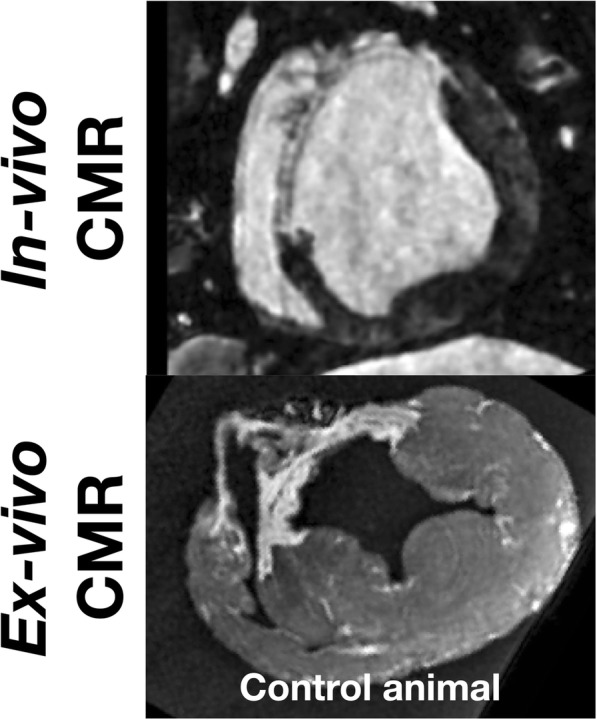
Ex-vivo cardiovascular magnetic resonance (CMR) imaging has played an important role in the validation of in-vivo CMR characterization of pathological processes. However, comparison between in-vivo and ex-vivo imaging remains challenging due to shape changes occurring between the two states, which may be non-uniform across the diseased heart. A novel two-step process to facilitate registration between ex-vivo and in-vivo CMR was developed and evaluated in a porcine model of chronic myocardial infarction (MI).

METHODS

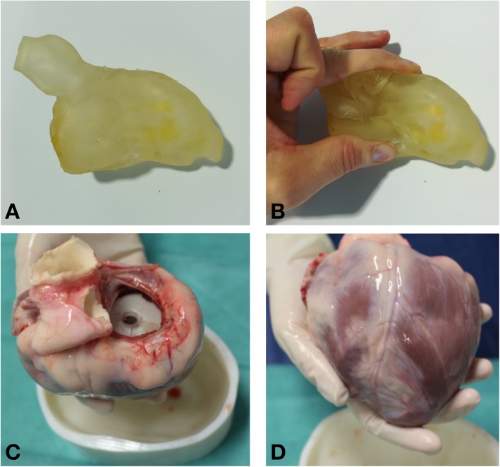
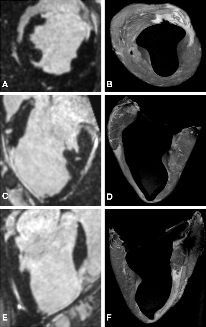
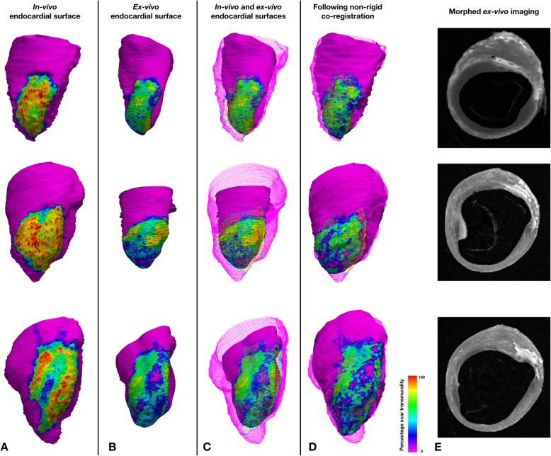
Seven weeks after ischemia-reperfusion MI, 12 swine underwent in-vivo CMR imaging with late gadolinium enhancement followed by ex-vivo CMR 1 week later. Five animals comprised the control group, in which ex-vivo imaging was undertaken without any support in the LV cavity, 7 animals comprised the experimental group, in which a two-step registration optimization process was undertaken. The first step involved a heart specific flexible 3D printed scaffold generated from in-vivo CMR, which was used to maintain left ventricular (LV) shape during ex-vivo imaging. In the second step, a non-rigid co-registration algorithm was applied to align in-vivo and ex-vivo data. Tissue dimension changes between in-vivo and ex-vivo imaging were compared between the experimental and control group. In the experimental group, tissue compartment volumes and thickness were compared between in-vivo and ex-vivo data before and after non-rigid registration. The effectiveness of the alignment was assessed quantitatively using the DICE similarity coefficient.

RESULTS

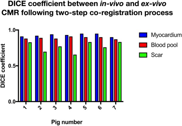
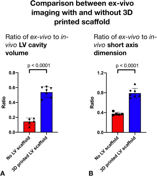
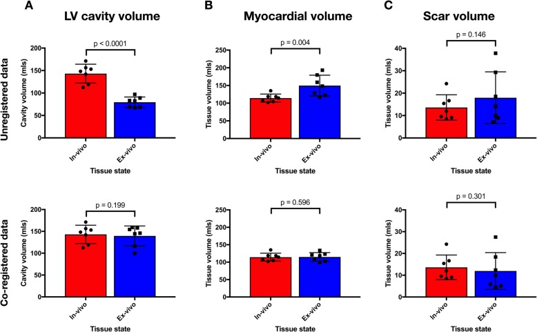
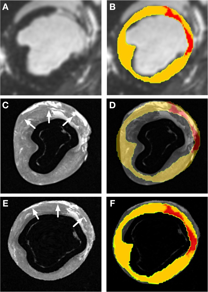
LV cavity volume changed more in the control group (ratio of cavity volume between ex-vivo and in-vivo imaging in control and experimental group 0.14 vs 0.56, p < 0.0001) and there was a significantly greater change in the short axis dimensions in the control group (ratio of short axis dimensions in control and experimental group 0.38 vs 0.79, p < 0.001). In the experimental group, prior to non-rigid co-registration the LV cavity contracted isotropically in the ex-vivo condition by less than 20% in each dimension. There was a significant proportional change in tissue thickness in the healthy myocardium (change = 29 ± 21%), but not in dense scar (change = - 2 ± 2%, p = 0.034). Following the non-rigid co-registration step of the process, the DICE similarity coefficients for the myocardium, LV cavity and scar were 0.93 (±0.02), 0.89 (±0.01) and 0.77 (±0.07) respectively and the myocardial tissue and LV cavity volumes had a ratio of 1.03 and 1.00 respectively.

CONCLUSIONS

The pattern of the morphological changes seen between the in-vivo and the ex-vivo LV differs between scar and healthy myocardium. A 3D printed flexible scaffold based on the in-vivo shape of the LV cavity is an effective strategy to minimize morphological changes in the ex-vivo LV. The subsequent non-rigid registration step further improved the co-registration and local comparison between in-vivo and ex-vivo data.

摘要

背景

在活体心脏磁共振(CMR)特征描述的验证中,离体心血管磁共振成像发挥了重要作用。然而,由于在两个状态之间发生的形状变化,活体和离体成像之间的比较仍然具有挑战性,这种变化在患病心脏中可能不是均匀的。本研究提出并评估了一种新的两步法,以促进慢性心肌梗死(MI)猪模型的离体和活体 CMR 之间的配准。

方法

在缺血再灌注 MI 后 7 周,12 只猪接受了晚期钆增强的活体 CMR 成像,1 周后进行了离体 CMR 成像。对照组中,5 只动物未在 LV 腔中进行任何支撑的情况下进行离体成像,实验组中,7 只动物进行了两步优化配准。第一步是使用活体 CMR 生成特定于心脏的柔性 3D 打印支架,以在离体成像过程中保持 LV 形状。在第二步中,应用非刚性配准算法来对齐活体和离体数据。比较实验组和对照组之间活体和离体成像之间的组织尺寸变化。在实验组中,比较了非刚性配准前后活体和离体数据的组织容积和厚度。使用 DICE 相似性系数定量评估对齐效果。

结果

LV 腔体积在对照组中变化更大(对照组和实验组的腔体积比为 0.14 比 0.56,p<0.0001),对照组的短轴尺寸变化更大(对照组和实验组的短轴尺寸比为 0.38 比 0.79,p<0.001)。在实验组中,在非刚性配准之前,每个维度的 LV 腔在离体条件下的收缩幅度都不到 20%。健康心肌组织的厚度有明显的比例变化(变化为 29±21%),但致密瘢痕组织没有(变化为-2±2%,p=0.034)。在该过程的非刚性配准步骤之后,心肌、LV 腔和瘢痕的 DICE 相似系数分别为 0.93(±0.02)、0.89(±0.01)和 0.77(±0.07),心肌组织和 LV 腔体积的比例分别为 1.03 和 1.00。

结论

活体和离体 LV 之间的形态变化模式在瘢痕和健康心肌组织之间有所不同。基于 LV 腔的体内形状的 3D 打印柔性支架是最小化离体 LV 形态变化的有效策略。随后的非刚性配准步骤进一步改善了活体和离体数据之间的配准和局部比较。

https://cdn.ncbi.nlm.nih.gov/pmc/blobs/5414/6785908/1a4c63499610/12968_2019_574_Fig8_HTML.jpg
https://cdn.ncbi.nlm.nih.gov/pmc/blobs/5414/6785908/230e52cf80de/12968_2019_574_Fig1_HTML.jpg
https://cdn.ncbi.nlm.nih.gov/pmc/blobs/5414/6785908/6a318dd11354/12968_2019_574_Fig2_HTML.jpg
https://cdn.ncbi.nlm.nih.gov/pmc/blobs/5414/6785908/408673724a92/12968_2019_574_Fig3_HTML.jpg
https://cdn.ncbi.nlm.nih.gov/pmc/blobs/5414/6785908/344e39779a0e/12968_2019_574_Fig4_HTML.jpg
https://cdn.ncbi.nlm.nih.gov/pmc/blobs/5414/6785908/3acc66ea57b3/12968_2019_574_Fig5_HTML.jpg
https://cdn.ncbi.nlm.nih.gov/pmc/blobs/5414/6785908/fe9c3c1c631f/12968_2019_574_Fig6_HTML.jpg
https://cdn.ncbi.nlm.nih.gov/pmc/blobs/5414/6785908/64148d605c12/12968_2019_574_Fig7_HTML.jpg
https://cdn.ncbi.nlm.nih.gov/pmc/blobs/5414/6785908/1a4c63499610/12968_2019_574_Fig8_HTML.jpg

相似文献

1
Improved co-registration of ex-vivo and in-vivo cardiovascular magnetic resonance images using heart-specific flexible 3D printed acrylic scaffold combined with non-rigid registration.使用心脏特异性柔性 3D 打印丙烯酸支架结合非刚性配准,提高离体和在体心血管磁共振图像的配准精度。
J Cardiovasc Magn Reson. 2019 Oct 10;21(1):62. doi: 10.1186/s12968-019-0574-z.
2
Implications of bipolar voltage mapping and magnetic resonance imaging resolution in biventricular scar characterization after myocardial infarction.双极电压图和磁共振成像分辨率对心肌梗死后双心室瘢痕特征的影响。
Europace. 2019 Jan 1;21(1):163-174. doi: 10.1093/europace/euy192.
3
Submillimeter diffusion tensor imaging and late gadolinium enhancement cardiovascular magnetic resonance of chronic myocardial infarction.慢性心肌梗死的亚毫米扩散张量成像及延迟钆增强心血管磁共振成像
J Cardiovasc Magn Reson. 2017 Jan 11;19(1):9. doi: 10.1186/s12968-016-0317-3.
4
A systematic comparison of cardiovascular magnetic resonance and high resolution histological fibrosis quantification in a chronic porcine infarct model.慢性猪梗死模型中心血管磁共振成像与高分辨率组织学纤维化定量分析的系统比较
Int J Cardiovasc Imaging. 2017 Nov;33(11):1797-1807. doi: 10.1007/s10554-017-1187-y. Epub 2017 Jun 14.
5
Experimental validation of contrast-enhanced SSFP cine CMR for quantification of myocardium at risk in acute myocardial infarction.对比增强稳态自由进动电影磁共振成像用于急性心肌梗死中危险心肌定量的实验验证
J Cardiovasc Magn Reson. 2017 Jan 30;19(1):12. doi: 10.1186/s12968-017-0325-y.
6
Contrast-enhanced cardiac MRI is superior to non-contrast mapping to predict left ventricular remodeling at 6 months after acute myocardial infarction.对比增强心脏 MRI 优于非对比映射,可预测急性心肌梗死后 6 个月的左心室重构。
Eur Radiol. 2024 Mar;34(3):1863-1874. doi: 10.1007/s00330-023-10100-9. Epub 2023 Sep 4.
7
Cardiovascular Magnetic Resonance Relaxometry Predicts Regional Functional Outcome After Experimental Myocardial Infarction.心血管磁共振弛豫测量法可预测实验性心肌梗死后的局部功能转归。
Circ Cardiovasc Imaging. 2017 Aug;10(8). doi: 10.1161/CIRCIMAGING.116.006025.
8
Detection and quantification of myocardial reperfusion hemorrhage using T2*-weighted CMR.利用 T2*-加权 CMR 检测和定量心肌再灌注出血。
JACC Cardiovasc Imaging. 2011 Dec;4(12):1274-83. doi: 10.1016/j.jcmg.2011.08.016.
9
In vivo chronic myocardial infarction characterization by spin locked cardiovascular magnetic resonance.自旋锁定心血管磁共振对体内慢性心肌梗死的特征描述。
J Cardiovasc Magn Reson. 2012 Jun 15;14(1):37. doi: 10.1186/1532-429X-14-37.
10
Regional and temporal changes in left ventricular strain and stiffness in a porcine model of myocardial infarction.心肌梗死后猪模型左心室应变和僵硬度的区域性和时间性变化。
Am J Physiol Heart Circ Physiol. 2018 Oct 1;315(4):H958-H967. doi: 10.1152/ajpheart.00279.2018. Epub 2018 Jul 13.

引用本文的文献

1
Combination of high resolution MRI with 3D-printed needle guides for ex vivo myocardial biopsies.高分辨率 MRI 与 3D 打印针引导器结合用于离体心肌活检。
Sci Rep. 2024 Jan 5;14(1):606. doi: 10.1038/s41598-023-50943-2.
2
Additional coils mitigate elevated defibrillation threshold in right-sided implantable cardioverter defibrillator generator placement: a simulation study.右侧植入式心律转复除颤器发生器放置中增加线圈可降低除颤阈值:一项模拟研究。
Europace. 2023 Jun 2;25(6). doi: 10.1093/europace/euad146.
3
Optimization of anti-tachycardia pacing efficacy through scar-specific delivery and minimization of re-initiation: a virtual study on a cohort of infarcted porcine hearts.

本文引用的文献

1
Pre-procedural fit-testing of TAVR valves using parametric modeling and 3D printing.经皮主动脉瓣置换术(TAVR)瓣膜的术前适配测试:参数建模与 3D 打印技术的应用。
J Cardiovasc Comput Tomogr. 2019 Jan-Feb;13(1):21-30. doi: 10.1016/j.jcct.2018.09.007. Epub 2018 Oct 2.
2
3D printing and its applications in orthopaedic trauma: A technological marvel.3D打印及其在骨科创伤中的应用:一项技术奇迹。
J Clin Orthop Trauma. 2018 Jul-Sep;9(3):260-268. doi: 10.1016/j.jcot.2018.07.022. Epub 2018 Aug 3.
3
Role of 3-Dimensional Architecture of Scar and Surviving Tissue in Ventricular Tachycardia: Insights From High-Resolution Ex Vivo Porcine Models.
通过瘢痕特异性传递和最小化再启动来优化抗心动过速起搏效果:对一组梗死猪心的虚拟研究。
Europace. 2023 Feb 16;25(2):716-725. doi: 10.1093/europace/euac165.
4
An automated near-real time computational method for induction and treatment of scar-related ventricular tachycardias.一种用于诱导和治疗瘢痕相关室性心动过速的自动化近实时计算方法。
Med Image Anal. 2022 Aug;80:102483. doi: 10.1016/j.media.2022.102483. Epub 2022 May 27.
5
Determining anatomical and electrophysiological detail requirements for computational ventricular models of porcine myocardial infarction.确定猪心肌梗死计算心室模型的解剖和电生理细节要求。
Comput Biol Med. 2022 Feb;141:105061. doi: 10.1016/j.compbiomed.2021.105061. Epub 2021 Nov 26.
6
An in-silico assessment of efficacy of two novel intra-cardiac electrode configurations versus traditional anti-tachycardia pacing therapy for terminating sustained ventricular tachycardia.两种新型心内电极配置与传统抗心动过速起搏治疗终止持续性室性心动过速的疗效的计算机评估。
Comput Biol Med. 2021 Dec;139:104987. doi: 10.1016/j.compbiomed.2021.104987. Epub 2021 Oct 30.
7
CemrgApp: An interactive medical imaging application with image processing, computer vision, and machine learning toolkits for cardiovascular research.CemrgApp:一款用于心血管研究的交互式医学成像应用程序,具备图像处理、计算机视觉和机器学习工具包。
SoftwareX. 2020 Jul 31;12:100570. doi: 10.1016/j.softx.2020.100570. eCollection 2020 Jul-Dec.
8
Three-dimensional printing for cardiovascular diseases: from anatomical modeling to dynamic functionality.三维打印在心血管疾病中的应用:从解剖建模到动态功能。
Biomed Eng Online. 2020 Oct 7;19(1):76. doi: 10.1186/s12938-020-00822-y.
瘢痕和存活组织的三维结构在室性心动过速中的作用:来自高分辨率猪离体模型的见解。
Circ Arrhythm Electrophysiol. 2018 Jun;11(6):e006131. doi: 10.1161/CIRCEP.117.006131.
4
Dual-energy micro-CT for quantifying the time-course and staining characteristics of ex-vivo animal organs treated with iodine- and gadolinium-based contrast agents.双能 micro-CT 定量分析碘基和钆基对比剂处理的离体动物器官的时程和染色特征。
Sci Rep. 2017 Dec 12;7(1):17387. doi: 10.1038/s41598-017-17064-z.
5
Imaging-Based Simulations for Predicting Sudden Death and Guiding Ventricular Tachycardia Ablation.基于成像的模拟用于预测猝死及指导室性心动过速消融
Circ Arrhythm Electrophysiol. 2017 Jul;10(7). doi: 10.1161/CIRCEP.117.004743.
6
Comparison of and MRI for the Detection of Structural Abnormalities in a Mouse Model of Tauopathy.用于检测tau蛋白病小鼠模型结构异常的[具体内容]与MRI比较
Front Neuroinform. 2017 Mar 31;11:20. doi: 10.3389/fninf.2017.00020. eCollection 2017.
7
Improved Magnetic Resonance Imaging-Pathology Correlation With Imaging-Derived, 3D-Printed, Patient-Specific Whole-Mount Molds of the Prostate.通过成像衍生的、3D打印的、患者特异性前列腺全层模型改善磁共振成像与病理学的相关性。
Invest Radiol. 2017 Sep;52(9):507-513. doi: 10.1097/RLI.0000000000000372.
8
Optimization of Vascular Casting for Three-Dimensional Fluorescence Cryo-Imaging of Collateral Vessels in the Ischemic Rat Hindlimb.缺血大鼠后肢侧支血管三维荧光冷冻成像的血管铸型优化
Microsc Microanal. 2017 Feb;23(1):77-87. doi: 10.1017/S1431927617000095.
9
Experimental validation of contrast-enhanced SSFP cine CMR for quantification of myocardium at risk in acute myocardial infarction.对比增强稳态自由进动电影磁共振成像用于急性心肌梗死中危险心肌定量的实验验证
J Cardiovasc Magn Reson. 2017 Jan 30;19(1):12. doi: 10.1186/s12968-017-0325-y.
10
Submillimeter diffusion tensor imaging and late gadolinium enhancement cardiovascular magnetic resonance of chronic myocardial infarction.慢性心肌梗死的亚毫米扩散张量成像及延迟钆增强心血管磁共振成像
J Cardiovasc Magn Reson. 2017 Jan 11;19(1):9. doi: 10.1186/s12968-016-0317-3.