Liang Fan, Ren Chunxia, Wang Jingshu, Wang Shuoer, Yang Lina, Han Xianghui, Chen Yaping, Tong Guoqing, Yang Gong
Department of Obstetrics and Gynecology, The Fifth People's Hospital of Shanghai, Fudan University, Shanghai, 200240, China.
Central Laboratory, the Fifth People's Hospital of Shanghai, Fudan University, Shanghai, 200240, China.
Oncogenesis. 2019 Oct 9;8(10):59. doi: 10.1038/s41389-019-0165-8.
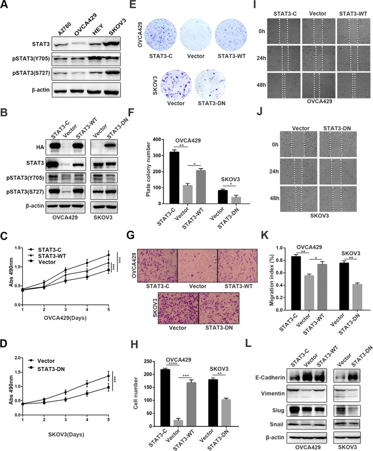
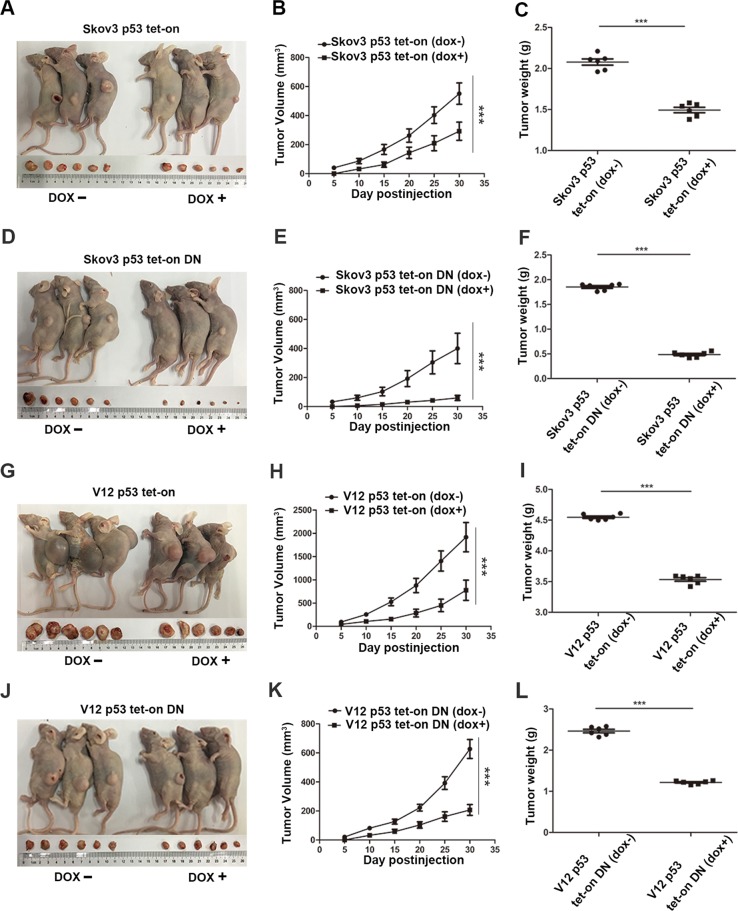
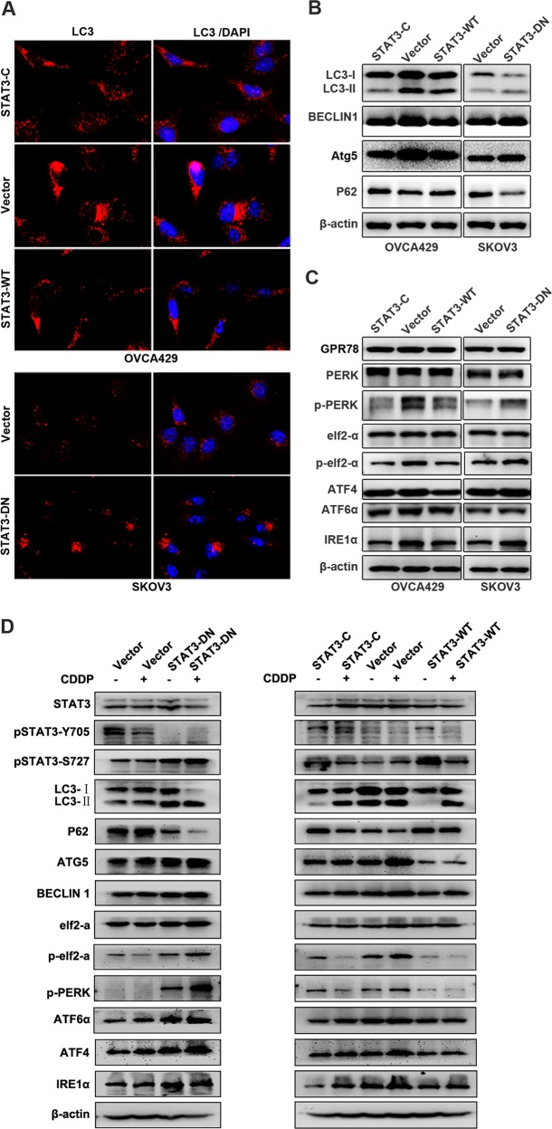
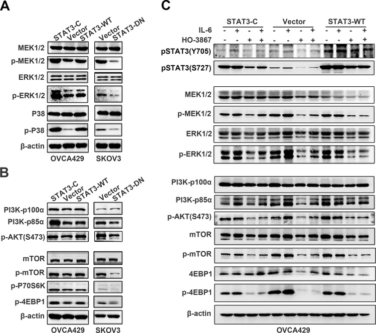
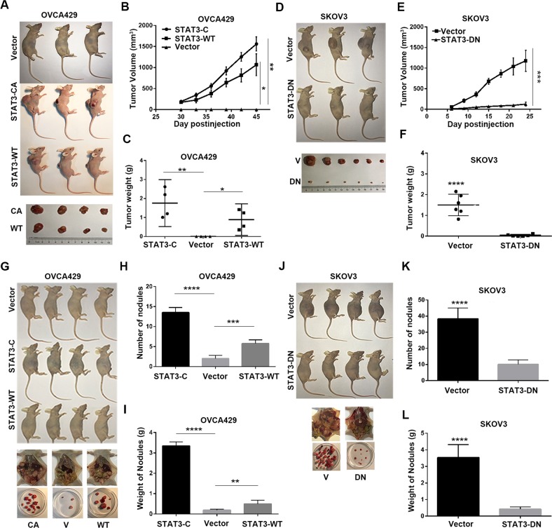
Chemoresistance has been the biggest obstacle in ovarian cancer treatment, and STAT3 may play an important role in chemoresistance of multiple cancers, but the underlying mechanism of STAT3 in ovarian cancer chemoresistance has long been truly illusive, particularly in association with p53 and RAS signaling. In this study, by using wild type, constitutive active, and dominant negative STAT3 constructs, wild-type p53, and RAS-mutant V12, we performed a series of in vitro and in vivo experiments by gene overexpression, drug treatment, and animal assays. We found that phosphorylation of STAT3 Y705 but not S727 promoted cancer cell EMT and metastasis through the Slug-mediated regulation of E-cadherin and Vimentin. The phosphorylation of STAT3 at Y705 also activated the MAPK and PI3K/AKT signaling to inhibit the ERS-mediated autophagy through down-regulation of pPERK, pelf2α, ATF6α, and IRE1α, which led to increased cisplatin resistance. Induction of wild type p53 in STAT3-DN-transfected cells further diminished the chemoresistance and tumor growth through the upregulation of the MAPK- and PI3K/AKT-mediated ERS and autophagy. Introduction of STAT3-DN deprived the RAS-induced ERS, autophagy, oncogenicity, and cisplatin resistance, whereas introduction of p53 in STAT3-DN/RAS expressing cells induced additional tumor retardation and cisplatin sensitivity. Thus, our data provide strong evidence that the crosstalk between STAT3 and p53/RAS signaling controls ovarian cancer cell metastasis and cisplatin resistance via the Slug/MAPK/PI3K/AKT-mediated regulation of EMT and autophagy.
化疗耐药一直是卵巢癌治疗中的最大障碍,STAT3可能在多种癌症的化疗耐药中发挥重要作用,但STAT3在卵巢癌化疗耐药中的潜在机制长期以来一直十分模糊,尤其是与p53和RAS信号相关的机制。在本研究中,我们使用野生型、组成型激活型和显性负性STAT3构建体、野生型p53以及RAS突变体V12,通过基因过表达、药物处理和动物实验进行了一系列体外和体内实验。我们发现,STAT3 Y705而非S727的磷酸化通过Slug介导的E-钙黏蛋白和波形蛋白调节促进癌细胞上皮-间质转化(EMT)和转移。STAT3在Y705位点的磷酸化还激活了MAPK和PI3K/AKT信号,通过下调pPERK、pelf2α、ATF6α和IRE1α来抑制内质网应激(ERS)介导的自噬,从而导致顺铂耐药性增加。在转染了STAT3显性负性构建体的细胞中诱导野生型p53表达,可通过上调MAPK和PI3K/AKT介导的ERS和自噬,进一步降低化疗耐药性并抑制肿瘤生长。引入STAT3显性负性构建体可消除RAS诱导的ERS、自噬、致癌性和顺铂耐药性,而在表达STAT3显性负性构建体/RAS的细胞中引入p53可诱导额外的肿瘤生长抑制和顺铂敏感性。因此,我们的数据提供了强有力证据,表明STAT3与p53/RAS信号之间的相互作用通过Slug/MAPK/PI3K/AKT介导的EMT和自噬调节,控制卵巢癌细胞的转移和顺铂耐药性。