Media Lab, MIT, Cambridge, MA, USA.
MIT McGovern Institute for Brain Research, MIT, Cambridge, MA, USA.
Nature. 2019 Oct;574(7778):413-417. doi: 10.1038/s41586-019-1641-1. Epub 2019 Oct 9.
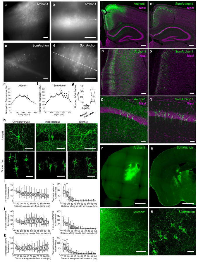
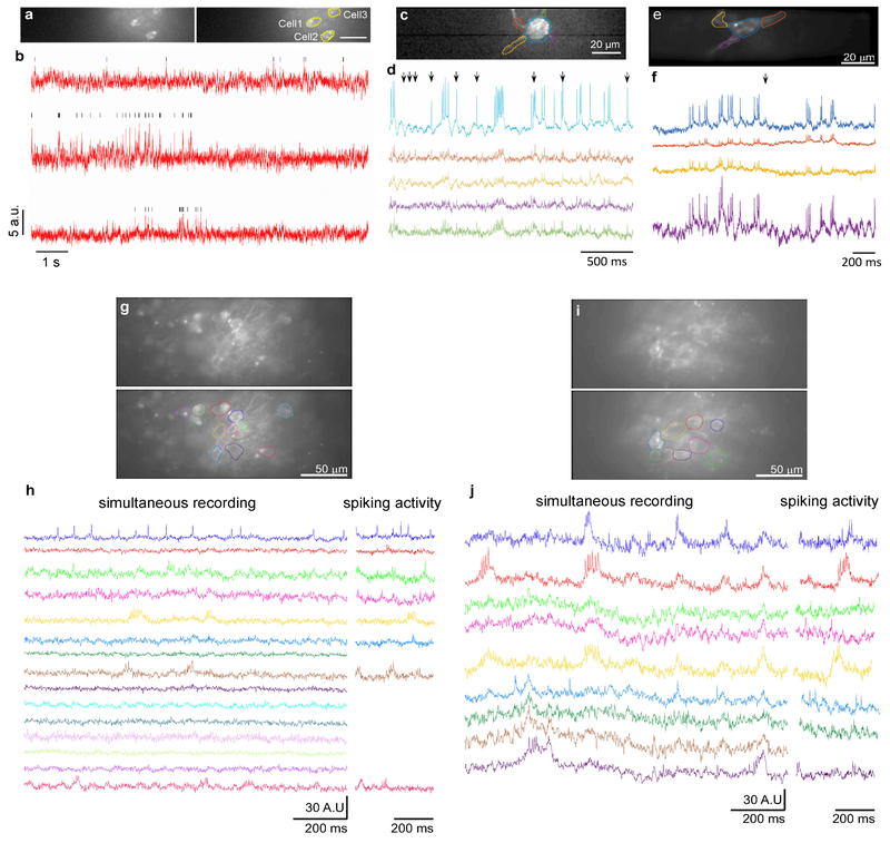
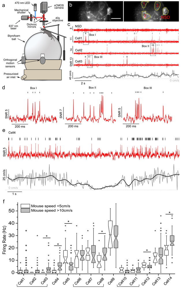
A longstanding goal in neuroscience has been to image membrane voltage across a population of individual neurons in an awake, behaving mammal. Here we describe a genetically encoded fluorescent voltage indicator, SomArchon, which exhibits millisecond response times and is compatible with optogenetic control, and which increases the sensitivity, signal-to-noise ratio, and number of neurons observable several-fold over previously published fully genetically encoded reagents. Under conventional one-photon microscopy, SomArchon enables the routine population analysis of around 13 neurons at once, in multiple brain regions (cortex, hippocampus, and striatum) of head-fixed, awake, behaving mice. Using SomArchon, we detected both positive and negative responses of striatal neurons during movement, as previously reported by electrophysiology but not easily detected using modern calcium imaging techniques, highlighting the power of voltage imaging to reveal bidirectional modulation. We also examined how spikes relate to the subthreshold theta oscillations of individual hippocampal neurons, with SomArchon showing that the spikes of individual neurons are more phase-locked to their own subthreshold theta oscillations than to local field potential theta oscillations. Thus, SomArchon reports both spikes and subthreshold voltage dynamics in awake, behaving mice.
在神经科学中,一个长期目标是在清醒、行为正常的哺乳动物中对单个神经元群体的膜电压进行成像。在这里,我们描述了一种遗传编码的荧光电压指示剂 SomArchon,它具有毫秒级的响应时间,与光遗传学控制兼容,并且与以前发表的完全遗传编码试剂相比,提高了灵敏度、信噪比和可观察神经元的数量数倍。在传统的单光子显微镜下,SomArchon 能够同时对 13 个左右的神经元进行常规的群体分析,这些神经元位于头部固定、清醒、行为正常的小鼠的多个脑区(皮层、海马体和纹状体)。使用 SomArchon,我们检测到运动过程中纹状体神经元的正负反应,这与电生理学的先前报道一致,但使用现代钙成像技术不易检测到,这突出了电压成像揭示双向调制的能力。我们还研究了单个海马体神经元的亚阈值 theta 振荡与尖峰之间的关系,SomArchon 表明单个神经元的尖峰与其自身亚阈值 theta 振荡的相位锁定程度高于局部场电势 theta 振荡。因此,SomArchon 在清醒、行为正常的小鼠中报告了尖峰和亚阈值电压动力学。