Department of Biology, Stanford University, Stanford, CA 94305.
Mol Biol Cell. 2019 Nov 15;30(24):2985-2995. doi: 10.1091/mbc.E19-03-0171. Epub 2019 Oct 10.
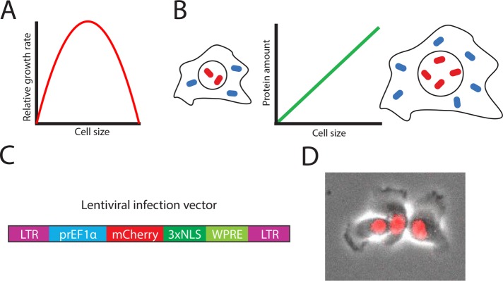
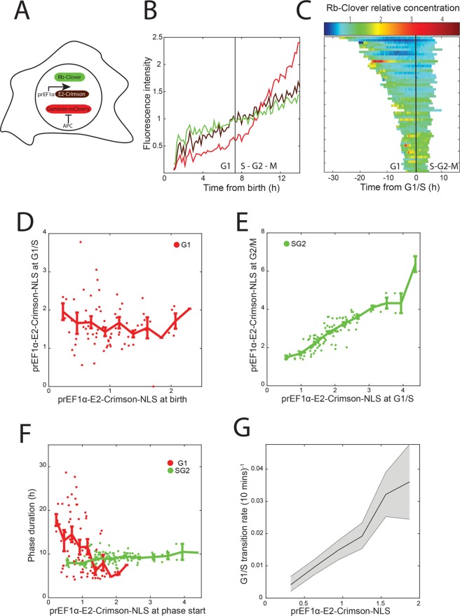
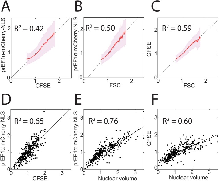
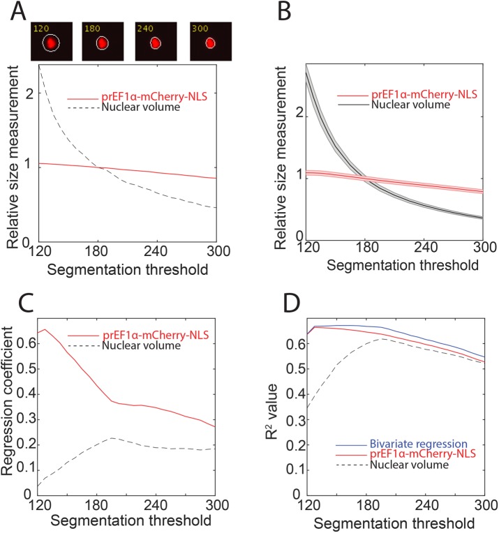
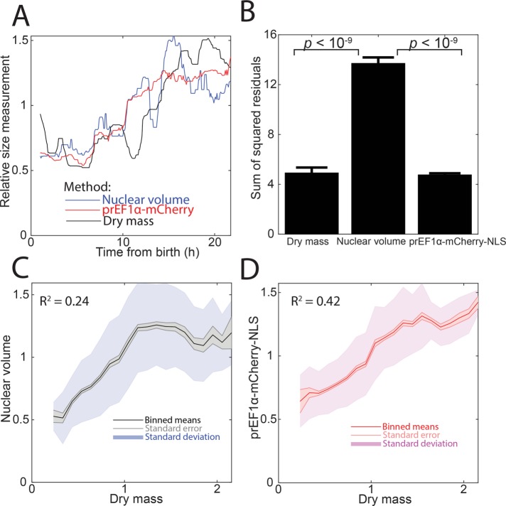
Cell size is important for cell physiology because it sets the geometric scale of organelles and biosynthesis. A number of methods exist to measure different aspects of cell size, but each has significant drawbacks. Here, we present an alternative method to measure the size of single human cells using a nuclear localized fluorescent protein expressed from a constitutive promoter. We validate this method by comparing it to several established cell size measurement strategies, including flow cytometry optical scatter, total protein dyes, and quantitative phase microscopy. We directly compare our fluorescent protein measurement with the commonly used measurement of nuclear volume and show that our measurements are more robust and less dependent on image segmentation. We apply our method to examine how cell size impacts the cell division cycle and reaffirm that there is a negative correlation between size at cell birth and G1 duration. Importantly, combining our size reporter with fluorescent labeling of a different protein in a different color channel allows measurement of concentration dynamics using simple wide-field fluorescence imaging. Thus, we expect our method will be of use to researchers interested in how dynamically changing protein concentrations control cell fates.
细胞大小对于细胞生理学很重要,因为它决定了细胞器和生物合成的几何尺度。有许多方法可以测量细胞大小的不同方面,但每种方法都有明显的缺点。在这里,我们提出了一种使用从组成型启动子表达的核定位荧光蛋白来测量单个人类细胞大小的替代方法。我们通过将其与几种已建立的细胞大小测量策略(包括流式细胞术光散射、总蛋白染料和定量相显微镜)进行比较来验证该方法。我们将我们的荧光蛋白测量与常用的核体积测量进行直接比较,并表明我们的测量更稳健,对图像分割的依赖性更小。我们应用我们的方法来研究细胞大小如何影响细胞分裂周期,并再次证实细胞出生时的大小与 G1 持续时间之间存在负相关。重要的是,将我们的大小报告基因与不同颜色通道中不同蛋白质的荧光标记相结合,允许使用简单的宽场荧光成像测量浓度动力学。因此,我们预计我们的方法将对研究动态变化的蛋白质浓度如何控制细胞命运的研究人员有用。