• 文献检索
  • 文档翻译
  • 深度研究
  • 学术资讯
  • Suppr Zotero 插件Zotero 插件
  • 邀请有礼
  • 套餐&价格
  • 历史记录
应用&插件
Suppr Zotero 插件Zotero 插件浏览器插件Mac 客户端Windows 客户端微信小程序
定价
高级版会员购买积分包购买API积分包
服务
文献检索文档翻译深度研究API 文档MCP 服务
关于我们
关于 Suppr公司介绍联系我们用户协议隐私条款
关注我们

Suppr 超能文献

核心技术专利:CN118964589B侵权必究
粤ICP备2023148730 号-1Suppr @ 2026

文献检索

告别复杂PubMed语法,用中文像聊天一样搜索,搜遍4000万医学文献。AI智能推荐,让科研检索更轻松。

立即免费搜索

文件翻译

保留排版,准确专业,支持PDF/Word/PPT等文件格式,支持 12+语言互译。

免费翻译文档

深度研究

AI帮你快速写综述,25分钟生成高质量综述,智能提取关键信息,辅助科研写作。

立即免费体验

一种课程学习策略,用于提高胸部 PA 射线筛查中各种病变分类的准确性,以发现肺部异常。

A Curriculum Learning Strategy to Enhance the Accuracy of Classification of Various Lesions in Chest-PA X-ray Screening for Pulmonary Abnormalities.

机构信息

University of Ulsan College of Medicine, Asan Medical Center, Department of Convergence Medicine, Seoul, South Korea.

University of Ulsan College of Medicine, Asan Medical Center, Department of Radiology and Research Institute of Radiology, Seoul, Korea.

出版信息

Sci Rep. 2019 Oct 25;9(1):15352. doi: 10.1038/s41598-019-51832-3.

DOI:10.1038/s41598-019-51832-3
PMID:31653943
原文链接:https://pmc.ncbi.nlm.nih.gov/articles/PMC6814828/
Abstract

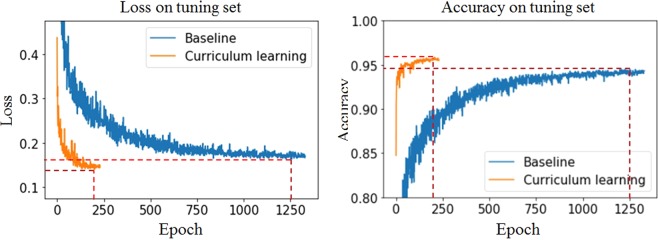
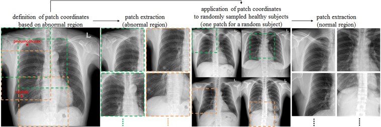
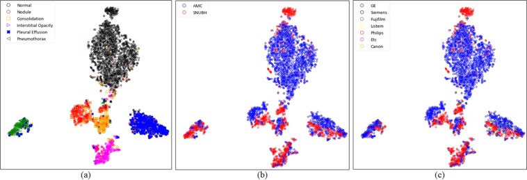
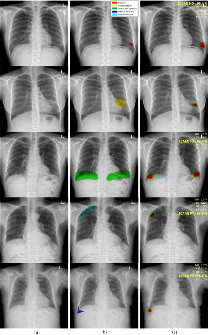
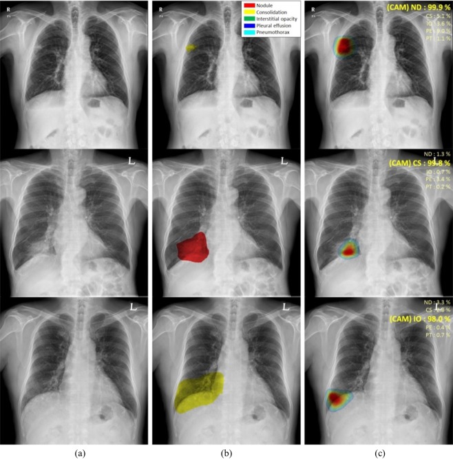
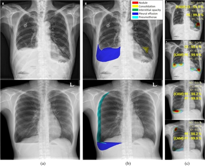
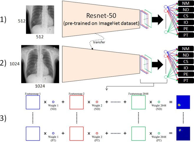
We evaluated the efficacy of a curriculum learning strategy using two steps to detect pulmonary abnormalities including nodule[s], consolidation, interstitial opacity, pleural effusion, and pneumothorax with chest-PA X-ray (CXR) images from two centres. CXR images of 6069 healthy subjects and 3417 patients at AMC and 1035 healthy subjects and 4404 patients at SNUBH were obtained. Our approach involved two steps. First, the regional patterns of thoracic abnormalities were identified by initial learning of patch images around abnormal lesions. Second, Resnet-50 was fine-tuned using the entire images. The network was weakly trained and modified to detect various disease patterns. Finally, class activation maps (CAM) were extracted to localise and visualise the abnormal patterns. For average disease, the sensitivity, specificity, and area under the curve (AUC) were 85.4%, 99.8%, and 0.947, respectively, in the AMC dataset and 97.9%, 100.0%, and 0.983, respectively, in the SNUBH dataset. This curriculum learning and weak labelling with high-scale CXR images requires less preparation to train the system and could be easily extended to include various diseases in actual clinical environments. This algorithm performed well for the detection and classification of five disease patterns in CXR images and could be helpful in image interpretation.

摘要

我们评估了一种使用两步法的课程学习策略的疗效,该策略用于从两个中心的胸部 PA X 射线(CXR)图像中检测肺异常,包括结节、实变、间质混浊、胸腔积液和气胸。我们的方法涉及两个步骤。首先,通过对异常病变周围的斑块图像进行初步学习,确定胸部异常的区域模式。其次,使用整个图像对 Resnet-50 进行微调。该网络进行了弱训练和修改,以检测各种疾病模式。最后,提取类激活图(CAM)以定位和可视化异常模式。对于常见疾病,在 AMC 数据集的灵敏度、特异性和曲线下面积(AUC)分别为 85.4%、99.8%和 0.947,在 SNUBH 数据集的灵敏度、特异性和 AUC 分别为 97.9%、100.0%和 0.983。这种使用大规模 CXR 图像的课程学习和弱标签需要较少的准备来训练系统,并且可以轻松扩展到实际临床环境中的各种疾病。该算法在 CXR 图像中五种疾病模式的检测和分类方面表现良好,有助于图像解释。

https://cdn.ncbi.nlm.nih.gov/pmc/blobs/d425/6814828/049b1ac099c5/41598_2019_51832_Fig7_HTML.jpg
https://cdn.ncbi.nlm.nih.gov/pmc/blobs/d425/6814828/13d0672a9f2d/41598_2019_51832_Fig1_HTML.jpg
https://cdn.ncbi.nlm.nih.gov/pmc/blobs/d425/6814828/11e8308b5cf2/41598_2019_51832_Fig2_HTML.jpg
https://cdn.ncbi.nlm.nih.gov/pmc/blobs/d425/6814828/c05aac127e5d/41598_2019_51832_Fig3_HTML.jpg
https://cdn.ncbi.nlm.nih.gov/pmc/blobs/d425/6814828/54d752ef16ed/41598_2019_51832_Fig4_HTML.jpg
https://cdn.ncbi.nlm.nih.gov/pmc/blobs/d425/6814828/1e7c7fa35e01/41598_2019_51832_Fig5_HTML.jpg
https://cdn.ncbi.nlm.nih.gov/pmc/blobs/d425/6814828/7112ba451595/41598_2019_51832_Fig6_HTML.jpg
https://cdn.ncbi.nlm.nih.gov/pmc/blobs/d425/6814828/049b1ac099c5/41598_2019_51832_Fig7_HTML.jpg

相似文献

1
A Curriculum Learning Strategy to Enhance the Accuracy of Classification of Various Lesions in Chest-PA X-ray Screening for Pulmonary Abnormalities.一种课程学习策略,用于提高胸部 PA 射线筛查中各种病变分类的准确性,以发现肺部异常。
Sci Rep. 2019 Oct 25;9(1):15352. doi: 10.1038/s41598-019-51832-3.
2
Optimal number of strong labels for curriculum learning with convolutional neural network to classify pulmonary abnormalities in chest radiographs.利用卷积神经网络进行课程学习以分类胸片肺部异常的最佳强标签数量。
Comput Biol Med. 2021 Sep;136:104750. doi: 10.1016/j.compbiomed.2021.104750. Epub 2021 Aug 9.
3
Automatic creation of annotations for chest radiographs based on the positional information extracted from radiographic image reports.基于从放射影像报告中提取的位置信息,为胸部 X 光片自动创建注释。
Comput Methods Programs Biomed. 2021 Sep;209:106331. doi: 10.1016/j.cmpb.2021.106331. Epub 2021 Aug 4.
4
High-precision multiclass classification of lung disease through customized MobileNetV2 from chest X-ray images.通过定制的MobileNetV2从胸部X光图像实现肺部疾病的高精度多类别分类。
Comput Biol Med. 2023 Mar;155:106646. doi: 10.1016/j.compbiomed.2023.106646. Epub 2023 Feb 10.
5
Deep transfer learning to quantify pleural effusion severity in chest X-rays.深度学习在胸部 X 光片中量化胸腔积液严重程度。
BMC Med Imaging. 2022 May 27;22(1):100. doi: 10.1186/s12880-022-00827-0.
6
A disentangled generative model for disease decomposition in chest X-rays via normal image synthesis.通过正常图像合成对胸部 X 光片中的疾病进行分解的解缠生成模型。
Med Image Anal. 2021 Jan;67:101839. doi: 10.1016/j.media.2020.101839. Epub 2020 Oct 7.
7
Multi-View Ensemble Convolutional Neural Network to Improve Classification of Pneumonia in Low Contrast Chest X-Ray Images.多视图集成卷积神经网络用于改善低对比度胸部X光图像中肺炎的分类
Annu Int Conf IEEE Eng Med Biol Soc. 2020 Jul;2020:1238-1241. doi: 10.1109/EMBC44109.2020.9176517.
8
Deep learning, reusable and problem-based architectures for detection of consolidation on chest X-ray images.深度学习,可复用且基于问题的架构,用于检测胸部 X 射线图像中的实变。
Comput Methods Programs Biomed. 2020 Mar;185:105162. doi: 10.1016/j.cmpb.2019.105162. Epub 2019 Oct 31.
9
Enhancing thoracic disease detection using chest X-rays from PubMed Central Open Access.利用 PubMed Central 开放获取中的胸部 X 光片增强胸部疾病检测。
Comput Biol Med. 2023 Jun;159:106962. doi: 10.1016/j.compbiomed.2023.106962. Epub 2023 Apr 20.
10
Proposing a novel multi-instance learning model for tuberculosis recognition from chest X-ray images based on CNNs, complex networks and stacked ensemble.提出了一种基于 CNNs、复杂网络和堆叠集成的新型多实例学习模型,用于从胸部 X 射线图像中识别肺结核。
Phys Eng Sci Med. 2021 Mar;44(1):291-311. doi: 10.1007/s13246-021-00980-w. Epub 2021 Feb 22.

引用本文的文献

1
Deep learning model for differentiating nasal cavity masses based on nasal endoscopy images.基于鼻内窥镜图像的鼻腔肿块深度学习模型。
BMC Med Inform Decis Mak. 2024 May 29;24(1):145. doi: 10.1186/s12911-024-02517-z.
2
Image Turing test and its applications on synthetic chest radiographs by using the progressive growing generative adversarial network.基于渐进式增长生成对抗网络的合成胸部 X 线图像图灵测试及其应用。
Sci Rep. 2023 Feb 9;13(1):2356. doi: 10.1038/s41598-023-28175-1.
3
Automatic lung segmentation in chest X-ray images using improved U-Net.

本文引用的文献

1
Deep Learning at Chest Radiography: Automated Classification of Pulmonary Tuberculosis by Using Convolutional Neural Networks.胸部放射摄影中的深度学习:使用卷积神经网络自动分类肺结核。
Radiology. 2017 Aug;284(2):574-582. doi: 10.1148/radiol.2017162326. Epub 2017 Apr 24.
2
Training and Validating a Deep Convolutional Neural Network for Computer-Aided Detection and Classification of Abnormalities on Frontal Chest Radiographs.训练和验证用于胸部正位X光片异常的计算机辅助检测与分类的深度卷积神经网络
Invest Radiol. 2017 May;52(5):281-287. doi: 10.1097/RLI.0000000000000341.
利用改进的 U-Net 进行胸部 X 光图像的自动肺分割。
Sci Rep. 2022 May 23;12(1):8649. doi: 10.1038/s41598-022-12743-y.
4
Segmentation and classification on chest radiography: a systematic survey.胸部X光片的分割与分类:一项系统综述。
Vis Comput. 2023;39(3):875-913. doi: 10.1007/s00371-021-02352-7. Epub 2022 Jan 8.
5
A Course-Focused Dual Curriculum For Image Captioning.一种针对图像字幕的以课程为重点的双轨课程。
Proc IEEE Int Symp Biomed Imaging. 2021 Apr;2021:716-720. doi: 10.1109/ISBI48211.2021.9434055. Epub 2021 May 25.
6
Deep convolution neural networks to differentiate between COVID-19 and other pulmonary abnormalities on chest radiographs: Evaluation using internal and external datasets.利用深度卷积神经网络在胸部X光片上区分新冠肺炎与其他肺部异常:基于内部和外部数据集的评估
Int J Imaging Syst Technol. 2021 Sep;31(3):1087-1104. doi: 10.1002/ima.22595. Epub 2021 May 13.
7
A Curriculum Learning Based Approach to Captioning Ultrasound Images.一种基于课程学习的超声图像字幕生成方法。
Med Ultrasound Preterm Perinat Paediatr Image Anal (2020). 2020 Oct;12437:75-84. doi: 10.1007/978-3-030-60334-2_8. Epub 2020 Oct 1.
8
Reproducibility of abnormality detection on chest radiographs using convolutional neural network in paired radiographs obtained within a short-term interval.使用卷积神经网络在短期内获得的配对胸片中检测胸部 X 光片异常的可重复性。
Sci Rep. 2020 Oct 15;10(1):17417. doi: 10.1038/s41598-020-74626-4.
9
Deep Learning in Medical Imaging.医学成像中的深度学习
Neurospine. 2019 Dec;16(4):657-668. doi: 10.14245/ns.1938396.198. Epub 2019 Dec 31.