Minnerly Christopher, Bressler Steven L, Shokry Ibrahim M, Tao Rui
Charles E. Schmidt College of Medicine, Florida Atlantic University, Boca Raton, FL, USA.
FHE Health, Deerfield Beach, FL, USA.
J Addict. 2019 Sep 26;2019:8586153. doi: 10.1155/2019/8586153. eCollection 2019.
Noninvasive estimation of cortical activity aberrance may be a challenge but gives valuable clues of mental health in patients. The goal of the present study was to characterize specificity of electroencephalogram (EEG) electrodes used to assess spectral powers associated with mental health conditions of patients with opioid use disorder.
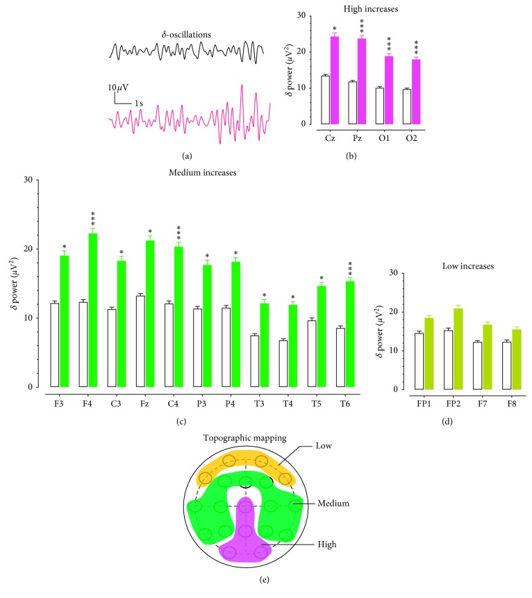
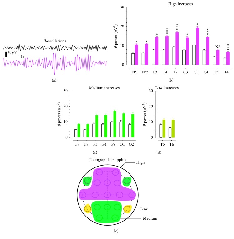
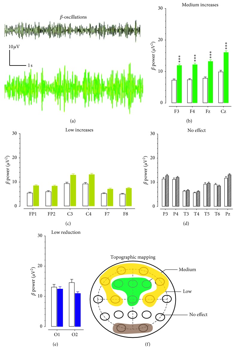
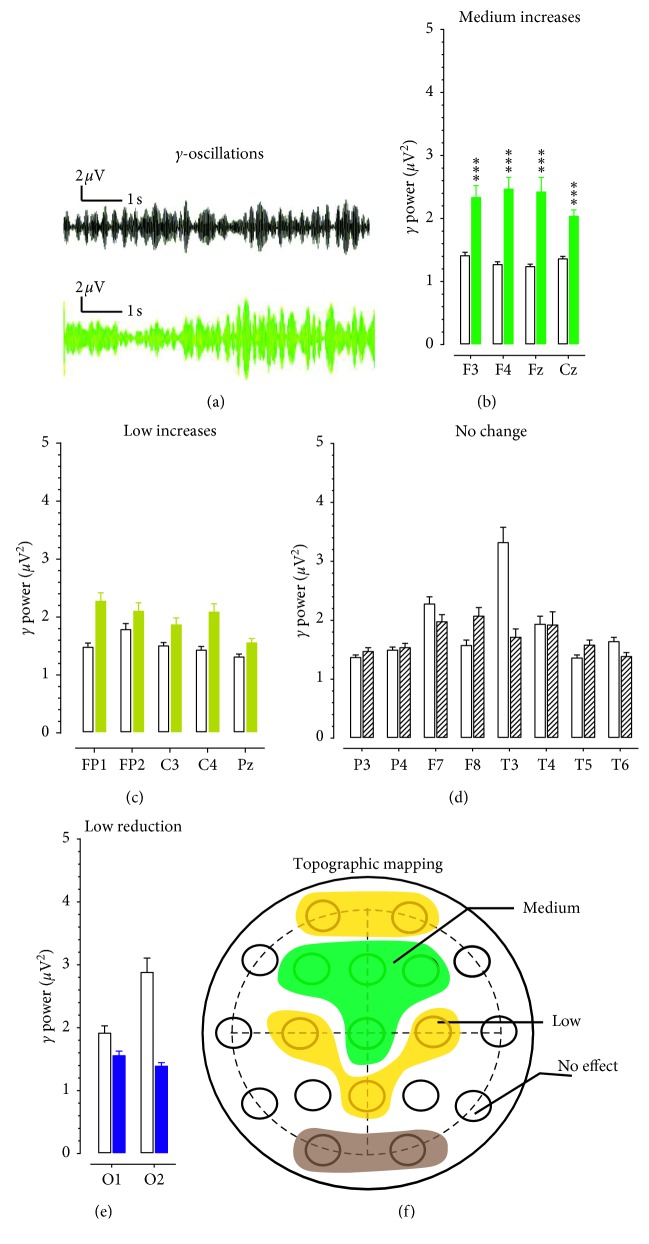
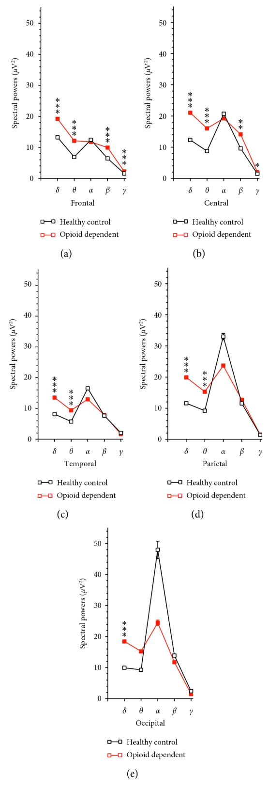
This retrospective study included 16 patients who had been diagnosed with opioid use disorder in comparison with 16 sex- and age-matched healthy controls. EEG electrodes were placed in the frontal (FP1, FP2, F3, F4, F7, F8, and Fz), central (C3, C4, and Cz), temporal (T3, T4, T5, and T6), parietal (P3, P4, and Pz), and occipital scalp (O1 and O2). Spectral powers of , , , , and γ oscillations were determined, and their distribution was topographically mapped with those electrodes on the scalp.
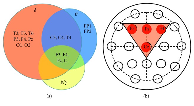
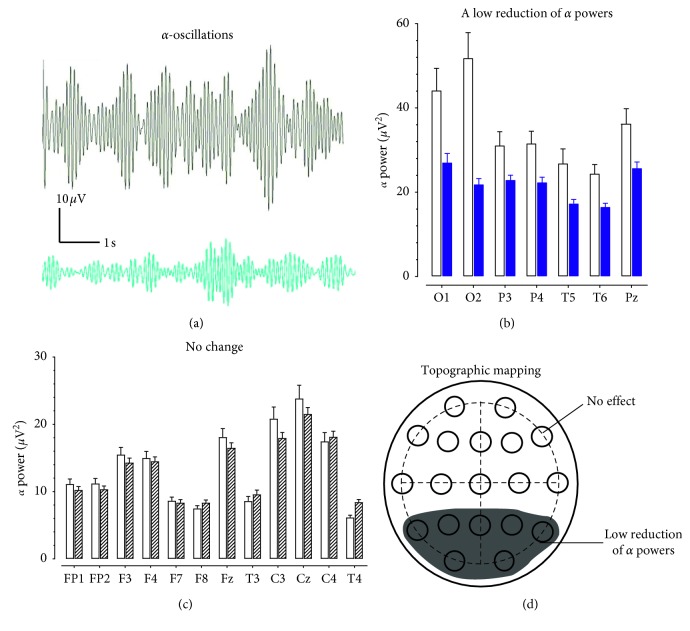
Compared to healthy controls, the spectral powers at low frequencies (<8 Hz; and ) were increased in most electrodes across the scalp, while powers at the high frequencies (>12 Hz; and γ) were selectively increased only at electrodes located in the frontal and central scalp. Among 19 electrodes, F3, F4, Fz, and Cz were highly specific in detecting increases in , , , and γ powers of patients with opioid use disorders.
Results of the present study demonstrate that spectral powers are topographically distributed across the scalp, which can be quantitatively characterized. Electrodes located at F3, F4, Fz, and Cz could be specifically utilized to assess mental health in patients with opioid use disorders. Mechanisms responsible for neuroplasticity involving cortical pyramidal neurons and -opioid receptor regulations are discussed within the context of changes in EEG microstates.
无创估计皮质活动异常可能具有挑战性,但能为患者的心理健康提供有价值的线索。本研究的目的是确定用于评估与阿片类物质使用障碍患者心理健康状况相关的频谱功率的脑电图(EEG)电极的特异性。
这项回顾性研究纳入了16例被诊断为阿片类物质使用障碍的患者,并与16例年龄和性别匹配的健康对照进行比较。EEG电极放置在额部(FP1、FP2、F3、F4、F7、F8和Fz)、中央(C3、C4和Cz)、颞部(T3、T4、T5和T6)、顶叶(P3、P4和Pz)以及枕部头皮(O1和O2)。测定了δ、θ、α、β和γ振荡的频谱功率,并使用头皮上的这些电极对其分布进行了地形图绘制。
与健康对照相比,头皮上大多数电极的低频(<8Hz;δ和θ)频谱功率增加,而高频(>12Hz;β和γ)功率仅在额部和中央头皮的电极上选择性增加。在19个电极中,F3、F4、Fz和Cz在检测阿片类物质使用障碍患者的δ、θ、β和γ功率增加方面具有高度特异性。
本研究结果表明,频谱功率在头皮上呈地形图分布,可以进行定量表征。位于F3、F4、Fz和Cz的电极可专门用于评估阿片类物质使用障碍患者的心理健康。在EEG微状态变化的背景下,讨论了涉及皮质锥体细胞和μ-阿片受体调节的神经可塑性的机制。