Department of Orthopaedic Surgery, The Third Hospital of Hebei Medical University, Shijiazhuang, China.
Department of Emergency Surgery, The First Hospital of Qinhuangdao Affiliated to Hebei Medical University, Qinhuangdao, China.
Orthop Surg. 2019 Oct;11(5):835-844. doi: 10.1111/os.12529.
To evaluate the dynamic changes of key morphology indicators of the lower extremities in the coronal plane with progressing medial compartment knee osteoarthritis (KOA) with an emphasis on gender-dependent regional differences.
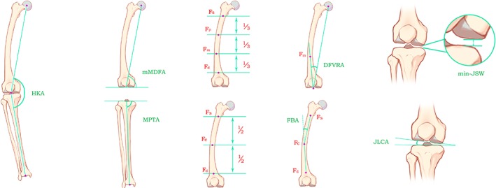
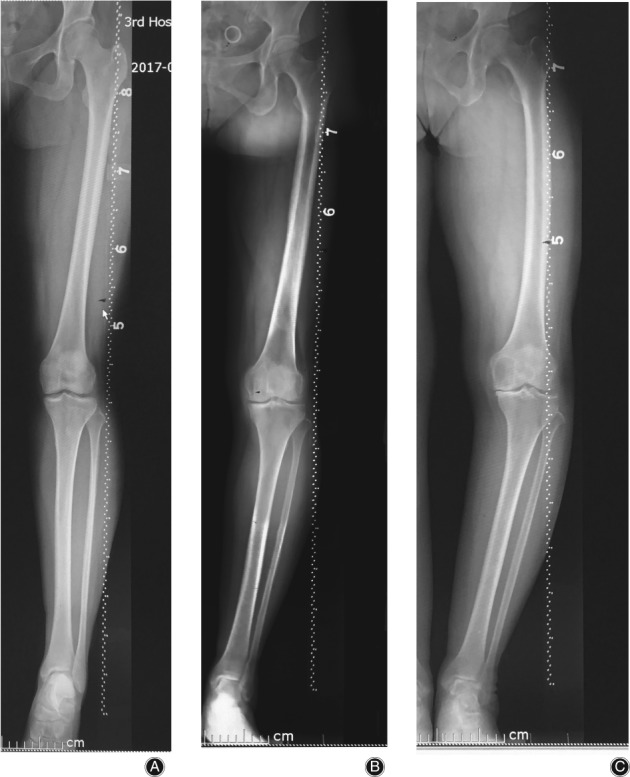
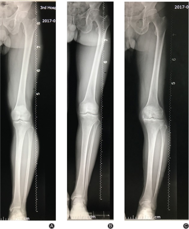
The radiographs of patients with non-traumatic knee pain and varying degrees of genu varus were reviewed. Radiographs were studied in 1538 lower limbs of 883 consecutive patients who visited our hospital from January to July 2017; all patients had long-standing anteroposterior image-splicing radiographs taken of their lower limbs. Morphological indicators of bones and joints that can change the alignment of lower limbs or reflect cartilage wear and soft-tissue relaxation were selected and measured with the help of picture archiving and communication systems. After comparing the data of different genders, the data of males and females was separated into three age groups, <40 years, 40-60 years, >60 years respectively, and then compared among age groups using the Kruskal-Wallis and Mann-Whitney U tests. Scatterplots of age and all the measurements were drawn to determine the strength of the relations. The Pearson correlation test was performed to reveal correlations of measurements and age.
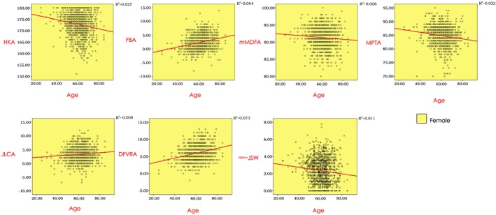
Femoral bowing angle (FBA) and joint line convergence angle (JLCA) have obvious differences between different genders (P = 0.001, 0.000, respectively). This suggests that females have greater femoral curvature and joint space angle than males. Significant differences were found in hip-knee-ankle angle (HKA), FBA, distal femoral valgus resection angle (DFVRA), medial proximal tibial angle (MPTA), JLCA, and minimum joint space width (min-JSW) by age groups in females (P = 0.000, 0.000, 0.000, 0.000, 0.003, 0.002, respectively). The difference of mechanical medial distal femoral angle (mMDFA) was significant with P values less than 0.05 deemed significant (P = 0.030). Significant correlations were found between age and all measurements (r = -0.166, 0.253, 0.270, -0.147, 0.089, -0.105, -0.076, respectively, P < 0.01). Whereas, the difference in min-JSW by age group was the only significant one in males (P = 0.001), and no significant correlation was found between age and measurements (r = -0.107, 0.041, 0.134, -0.067, 0.079, -0.134, -0.098, respectively, P > 0.01).
As KOA progressed, both dynamic deformation of lower extremities and degeneration of articular cartilage could be found in females, while no obvious dynamic deformations were found in males. Dynamic deformation of lower extremities was the important feature and the major causative factor of KOA in females.
评估进展性内侧间室膝骨关节炎(KOA)患者下肢冠状面关键形态指标的动态变化,重点关注性别相关的区域性差异。
回顾了非创伤性膝关节疼痛且存在不同程度内翻畸形的患者的影像学资料。2017 年 1 月至 7 月期间,对我院连续就诊的 883 例患者的 1538 条下肢的影像学资料进行了研究,所有患者均进行了下肢前后位图像拼接的影像学检查。选择了可能改变下肢对线或反映软骨磨损和软组织松弛的骨骼关节形态指标,并借助影像存档与通讯系统进行测量。在比较不同性别的数据后,将男性和女性的数据分别分为<40 岁、40-60 岁、>60 岁三个年龄组,然后使用 Kruskal-Wallis 和 Mann-Whitney U 检验对各年龄组进行比较。绘制年龄与所有测量值的散点图,以确定相关性的强弱。采用 Pearson 相关性检验揭示测量值与年龄之间的相关性。
不同性别间股骨弯曲角度(FBA)和关节线会聚角(JLCA)存在明显差异(P = 0.001,0.000)。这表明女性的股骨弯曲度和关节间隙角度大于男性。在女性中,HKA、FBA、远端股骨外翻切除角(DFVRA)、内侧胫骨近端角(MPTA)、JLCA 和最小关节间隙宽度(min-JSW)在不同年龄组间存在显著差异(P = 0.000,0.000,0.000,0.000,0.003,0.002)。机械性内侧远端股骨角(mMDFA)的差异具有统计学意义(P = 0.030)。年龄与所有测量值之间均存在显著相关性(r = -0.166,0.253,0.270,-0.147,0.089,-0.105,-0.076,均 P < 0.01)。而在男性中,仅 min-JSW 在年龄组间存在显著差异(P = 0.001),且年龄与测量值之间无显著相关性(r = -0.107,0.041,0.134,-0.067,0.079,-0.134,-0.098,均 P > 0.01)。
随着 KOA 的进展,女性患者可出现下肢的动态变形和关节软骨的退变,而男性患者则无明显的下肢动态变形。下肢的动态变形是女性 KOA 的重要特征和主要致病因素。