• 文献检索
  • 文档翻译
  • 深度研究
  • 学术资讯
  • Suppr Zotero 插件Zotero 插件
  • 邀请有礼
  • 套餐&价格
  • 历史记录
应用&插件
Suppr Zotero 插件Zotero 插件浏览器插件Mac 客户端Windows 客户端微信小程序
定价
高级版会员购买积分包购买API积分包
服务
文献检索文档翻译深度研究API 文档MCP 服务
关于我们
关于 Suppr公司介绍联系我们用户协议隐私条款
关注我们

Suppr 超能文献

核心技术专利:CN118964589B侵权必究
粤ICP备2023148730 号-1Suppr @ 2026

文献检索

告别复杂PubMed语法,用中文像聊天一样搜索,搜遍4000万医学文献。AI智能推荐,让科研检索更轻松。

立即免费搜索

文件翻译

保留排版,准确专业,支持PDF/Word/PPT等文件格式,支持 12+语言互译。

免费翻译文档

深度研究

AI帮你快速写综述,25分钟生成高质量综述,智能提取关键信息,辅助科研写作。

立即免费体验

糖尿病患者的视乳头周围微血管:一项光相干断层扫描血管造影研究。

Peripapillary microvasculature in patients with diabetes mellitus: An optical coherence tomography angiography study.

机构信息

Department of Ophthalmology, Chungnam National University College of Medicine, Daejeon, Republic of Korea.

Department of Ophthalmology, Gyeongsang National University Changwon Hospital, Changwon, Republic of Korea.

出版信息

Sci Rep. 2019 Nov 1;9(1):15814. doi: 10.1038/s41598-019-52354-8.

DOI:10.1038/s41598-019-52354-8
PMID:31676848
原文链接:https://pmc.ncbi.nlm.nih.gov/articles/PMC6825207/
Abstract

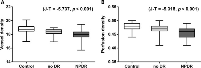
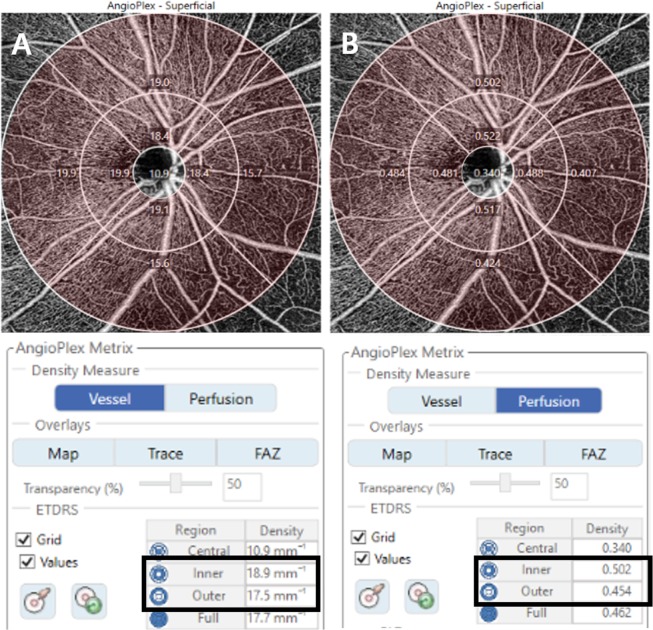
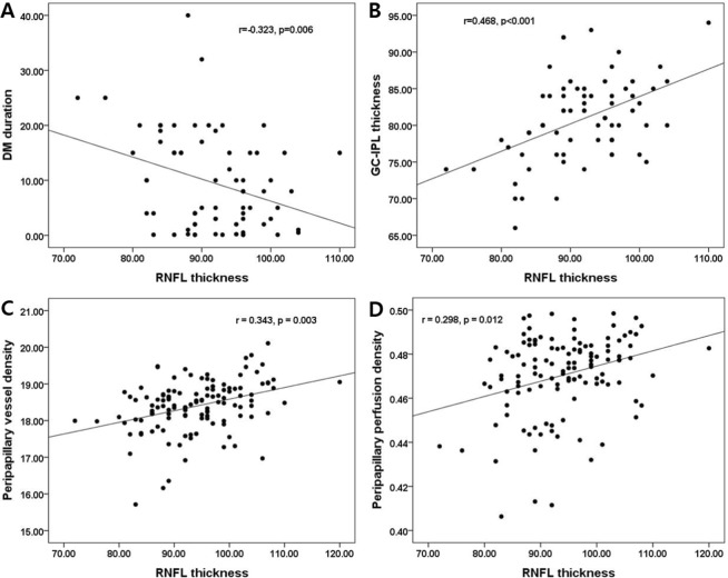
To evaluate changes in peripapillary microvascular parameters in diabetes mellitus (DM) patients using optical coherence tomography angiography (OCTA). Seventy-one diabetic patients (40 in the no diabetic retinopathy [DR] group and 31 in the non-proliferative DR [NPDR] group) and 50 control subjects. OCTA (Zeiss HD-OCT 5000 with AngioPlex) 6 × 6 mm scans centered on the optic disc were analyzed. Peripapillary vessel density (VD), perfusion density (PD) in superficial capillary plexus (SCP) were automatically calculated. The average macular ganglion cell-inner plexiform layer (mGC-IPL) and peripapillary retinal nerve fiber layer (pRNFL) thicknesses of the no DR and NPDR groups were significantly thinner than those of the control group. The no DR and NPDR groups showed lower peripapillary VD and PD in SCP compared with the control group. Using univariate regression analyses, the average mGC-IPL thickness, the pRNFL thickness, the no DR group and NPDR group were significant factors that affected the peripapillary VD and PD in SCP. Multivariate regression analyses showed that the grade of DR was a significant factor affecting the peripapillary VD and PD in SCP. OCTA revealed that peripapillary microvascular parameters in the no DR and NPDR groups were lower than those of normal controls. The peripapillary VD and PD in SCP were correlated with the mGC-IPL thickness, the pRNFL thickness, and the no DR and NPDR groups. Changes in peripapillary OCTA parameters may help with understanding the pathophysiology of DM and evaluating a potentially valuable biomarker for patients with subclinical DR.

摘要

利用光相干断层扫描血管造影术(OCTA)评估糖尿病患者的视盘周围微血管参数变化。共纳入 71 例糖尿病患者(无糖尿病视网膜病变[DR]组 40 例,非增生性 DR[NPDR]组 31 例)和 50 例对照者。对以视盘为中心的 6×6mm OCTA(蔡司 HD-OCT 5000 与 AngioPlex)进行分析。自动计算视盘周围血管密度(VD)和浅层毛细血管丛(SCP)的灌注密度(PD)。无 DR 组和 NPDR 组的平均黄斑神经节细胞-内丛状层(mGC-IPL)和视盘周围视网膜神经纤维层(pRNFL)厚度明显比对照组薄。与对照组相比,无 DR 组和 NPDR 组的视盘周围 VD 和 SCP 中的 PD 较低。采用单变量回归分析,平均 mGC-IPL 厚度、pRNFL 厚度、无 DR 组和 NPDR 组是影响 SCP 视盘周围 VD 和 PD 的显著因素。多变量回归分析显示,DR 分级是影响 SCP 视盘周围 VD 和 PD 的显著因素。OCTA 显示,无 DR 组和 NPDR 组的视盘周围微血管参数均低于正常对照组。SCP 中的视盘周围 VD 和 PD 与 mGC-IPL 厚度、pRNFL 厚度以及无 DR 组和 NPDR 组相关。视盘 OCTA 参数的变化可能有助于了解糖尿病的病理生理学,并评估亚临床 DR 患者有潜在价值的生物标志物。

https://cdn.ncbi.nlm.nih.gov/pmc/blobs/441c/6825207/3928ea8a848b/41598_2019_52354_Fig3_HTML.jpg
https://cdn.ncbi.nlm.nih.gov/pmc/blobs/441c/6825207/4722863c3182/41598_2019_52354_Fig1_HTML.jpg
https://cdn.ncbi.nlm.nih.gov/pmc/blobs/441c/6825207/92077be73009/41598_2019_52354_Fig2_HTML.jpg
https://cdn.ncbi.nlm.nih.gov/pmc/blobs/441c/6825207/3928ea8a848b/41598_2019_52354_Fig3_HTML.jpg
https://cdn.ncbi.nlm.nih.gov/pmc/blobs/441c/6825207/4722863c3182/41598_2019_52354_Fig1_HTML.jpg
https://cdn.ncbi.nlm.nih.gov/pmc/blobs/441c/6825207/92077be73009/41598_2019_52354_Fig2_HTML.jpg
https://cdn.ncbi.nlm.nih.gov/pmc/blobs/441c/6825207/3928ea8a848b/41598_2019_52354_Fig3_HTML.jpg

相似文献

1
Peripapillary microvasculature in patients with diabetes mellitus: An optical coherence tomography angiography study.糖尿病患者的视乳头周围微血管:一项光相干断层扫描血管造影研究。
Sci Rep. 2019 Nov 1;9(1):15814. doi: 10.1038/s41598-019-52354-8.
2
Changes in Peripapillary Microvasculature and Retinal Thickness in the Fellow Eyes of Patients With Unilateral Retinal Vein Occlusion: An OCTA Study.单侧视网膜静脉阻塞患者对侧眼的视盘周围微血管和视网膜厚度的变化:OCTA 研究。
Invest Ophthalmol Vis Sci. 2019 Feb 1;60(2):823-829. doi: 10.1167/iovs.18-26288.
3
Quantitative Analysis of the RPC Vessel Density and the RNFL Thickness in Patients with Type 2 Diabetes Mellitus by Using OCT Angiography.应用光学相干断层血管成像术定量分析 2 型糖尿病患者视网膜毛细血管密度和神经纤维层厚度。
Ophthalmic Res. 2021;64(6):951-959. doi: 10.1159/000517145. Epub 2021 Jul 20.
4
Macular Capillary Perfusion in Chinese Patients With Diabetic Retinopathy Obtained With Optical Coherence Tomography Angiography.光学相干断层扫描血管造影术检测中国糖尿病视网膜病变患者的黄斑区毛细血管灌注情况
Ophthalmic Surg Lasers Imaging Retina. 2019 Apr 1;50(4):e88-e95. doi: 10.3928/23258160-20190401-12.
5
Changes in Ganglion Cell-Inner Plexiform Layer Thickness and Retinal Microvasculature in Hypertension: An Optical Coherence Tomography Angiography Study.高血压患者神经节细胞-内丛状层厚度和视网膜微血管变化的光学相干断层血管造影研究。
Am J Ophthalmol. 2019 Mar;199:167-176. doi: 10.1016/j.ajo.2018.11.016. Epub 2018 Nov 29.
6
Peripapillary Retinal Nerve Fiber Layer and Microvasculature in Prolonged Type 2 Diabetes Patients Without Clinical Diabetic Retinopathy.无临床糖尿病视网膜病变的长期 2 型糖尿病患者的视乳头周围视网膜神经纤维层和微血管。
Invest Ophthalmol Vis Sci. 2021 Feb 1;62(2):9. doi: 10.1167/iovs.62.2.9.
7
Do microvascular changes occur preceding neural impairment in early-stage diabetic retinopathy? Evidence based on the optic nerve head using optical coherence tomography angiography.早期糖尿病性视网膜病变中,微血管改变是否先于神经损伤发生?基于视神经头的光相干断层扫描血管造影的证据。
Acta Diabetol. 2019 May;56(5):531-539. doi: 10.1007/s00592-019-01288-8. Epub 2019 Jan 17.
8
Peripapillary microvascular changes in patients with systemic hypertension: An optical coherence tomography angiography study.高血压患者视盘周围微血管变化的光学相干断层血管造影研究。
Sci Rep. 2020 Apr 16;10(1):6541. doi: 10.1038/s41598-020-63603-6.
9
Retinal Microvascular and Neurodegenerative Changes in Alzheimer's Disease and Mild Cognitive Impairment Compared with Control Participants.与对照参与者相比,阿尔茨海默病和轻度认知障碍中的视网膜微血管和神经退行性变化
Ophthalmol Retina. 2019 Jun;3(6):489-499. doi: 10.1016/j.oret.2019.02.002. Epub 2019 Mar 11.
10
Peripapillary Microvascular and Neural Changes in Diabetes Mellitus: An OCT-Angiography Study.糖尿病患者视乳头周围微血管和神经变化的 OCT 血管造影研究。
Invest Ophthalmol Vis Sci. 2018 Oct 1;59(12):5074-5081. doi: 10.1167/iovs.18-24891.

引用本文的文献

1
Early detection of retinal and choroidal microvascular impairments in diabetic patients with myopia.近视糖尿病患者视网膜和脉络膜微血管损伤的早期检测
Front Cell Dev Biol. 2025 May 26;13:1609928. doi: 10.3389/fcell.2025.1609928. eCollection 2025.
2
New marker for the detection of pre-retinopathy in patients with type 1 diabetes mellitus: systemic immuno-inflammation index.1型糖尿病患者视网膜病变前期检测的新标志物:全身免疫炎症指数。
BMC Ophthalmol. 2025 May 19;25(1):296. doi: 10.1186/s12886-025-04138-0.
3
Peripapillary Bruch's membrane Opening-Minimum Rim Width (BMO-MRW) and microvascular changes in early diabetic retinopathy.

本文引用的文献

1
Changes in Peripapillary Microvasculature and Retinal Thickness in the Fellow Eyes of Patients With Unilateral Retinal Vein Occlusion: An OCTA Study.单侧视网膜静脉阻塞患者对侧眼的视盘周围微血管和视网膜厚度的变化:OCTA 研究。
Invest Ophthalmol Vis Sci. 2019 Feb 1;60(2):823-829. doi: 10.1167/iovs.18-26288.
2
Retinal Microvascular Change in Hypertension as measured by Optical Coherence Tomography Angiography.光学相干断层扫描血管造影术测量的高血压视网膜微血管变化。
Sci Rep. 2019 Jan 17;9(1):156. doi: 10.1038/s41598-018-36474-1.
3
Changes in Ganglion Cell-Inner Plexiform Layer Thickness and Retinal Microvasculature in Hypertension: An Optical Coherence Tomography Angiography Study.
视乳头周围脉络膜毛细血管层开口-最小边缘宽度(BMO-MRW)与早期糖尿病视网膜病变中的微血管变化
BMC Ophthalmol. 2025 Apr 28;25(1):257. doi: 10.1186/s12886-025-04077-w.
4
Structure-Function Associations Between Quantitative Contrast Sensitivity Function And Peripapillary Optical Coherence Tomography Angiography in Diabetic Retinopathy.糖尿病视网膜病变中定量对比敏感度函数与视乳头周围光学相干断层扫描血管造影之间的结构-功能关联
Invest Ophthalmol Vis Sci. 2025 Apr 1;66(4):69. doi: 10.1167/iovs.66.4.69.
5
Using blood biomarkers and ophthalmological indicators of optical coherence tomography and angiography for the diagnosis of fundus lesions in patients with diabetes mellitus.利用血液生物标志物以及光学相干断层扫描和血管造影的眼科指标诊断糖尿病患者的眼底病变。
Front Clin Diabetes Healthc. 2025 Apr 8;6:1499344. doi: 10.3389/fcdhc.2025.1499344. eCollection 2025.
6
Changes in peripapillary microvasculature and retinal nerve fibre layer in diabetes and diabetic retinopathy using optical coherence tomographic angiography: a community-based, cross-sectional study.利用光学相干断层扫描血管造影术观察糖尿病及糖尿病视网膜病变患者视乳头周围微血管和视网膜神经纤维层的变化:一项基于社区的横断面研究。
BMJ Open. 2025 Mar 29;15(3):e079572. doi: 10.1136/bmjopen-2023-079572.
7
Sub-Clinical Regional Macular Changes in Self-Reported Diabetic Subjects: Vascular and Retinal Layer Analysis Using Swept-Source OCT.自我报告的糖尿病患者的亚临床区域黄斑变化:使用扫频光学相干断层扫描进行血管和视网膜层分析
Diabetes Metab Syndr Obes. 2025 Feb 6;18:363-372. doi: 10.2147/DMSO.S504882. eCollection 2025.
8
Retinal Microvascular Changes in Association with Endothelial Glycocalyx Damage and Arterial Stiffness in Patients with Diabetes Mellitus Type 2: A Cross-Sectional Study in a Greek Population.2型糖尿病患者视网膜微血管变化与内皮糖萼损伤及动脉僵硬度的相关性:希腊人群的横断面研究
J Pers Med. 2024 Sep 19;14(9):995. doi: 10.3390/jpm14090995.
9
Study of the impact of the vascular systemic risk factors on peripapillary vascular density by optical coherence tomography angiography.应用光相干断层扫描血管造影术研究血管系统危险因素对视盘周围血管密度的影响。
Graefes Arch Clin Exp Ophthalmol. 2024 Dec;262(12):3933-3946. doi: 10.1007/s00417-024-06576-w. Epub 2024 Jul 11.
10
Repeatability, interocular correlation and agreement of optic nerve head vessel density in healthy eyes: a swept-source optical coherence tomographic angiography study.健康眼睛视神经乳头血管密度的可重复性、眼间相关性及一致性:一项扫频源光学相干断层血管造影研究
Int J Ophthalmol. 2024 May 18;17(5):896-903. doi: 10.18240/ijo.2024.05.14. eCollection 2024.
高血压患者神经节细胞-内丛状层厚度和视网膜微血管变化的光学相干断层血管造影研究。
Am J Ophthalmol. 2019 Mar;199:167-176. doi: 10.1016/j.ajo.2018.11.016. Epub 2018 Nov 29.
4
Peripapillary Microvascular and Neural Changes in Diabetes Mellitus: An OCT-Angiography Study.糖尿病患者视乳头周围微血管和神经变化的 OCT 血管造影研究。
Invest Ophthalmol Vis Sci. 2018 Oct 1;59(12):5074-5081. doi: 10.1167/iovs.18-24891.
5
PERIPAPILLARY NEUROVASCULAR COUPLING IN THE EARLY STAGES OF DIABETIC RETINOPATHY.糖尿病性视网膜病变早期的视盘周围神经血管耦合。
Retina. 2019 Dec;39(12):2292-2302. doi: 10.1097/IAE.0000000000002328.
6
Optic nerve head perfusion changes preceding peripapillary retinal nerve fibre layer thinning in preclinical diabetic retinopathy.在临床前期糖尿病性视网膜病变中,视乳头头部灌注变化先于视乳头周围视网膜神经纤维层变薄。
Clin Exp Ophthalmol. 2019 Mar;47(2):219-225. doi: 10.1111/ceo.13390. Epub 2018 Nov 13.
7
The Importance of Signal Strength in Quantitative Assessment of Retinal Vessel Density Using Optical Coherence Tomography Angiography.光学相干断层扫描血管造影定量评估视网膜血管密度中信号强度的重要性。
Sci Rep. 2018 Aug 27;8(1):12897. doi: 10.1038/s41598-018-31321-9.
8
Optical coherence tomography angiography analysis of foveal microvascular changes and inner retinal layer thinning in patients with diabetes.糖尿病患者黄斑区微血管改变及内层视网膜变薄的光相干断层扫描血管造影分析。
Br J Ophthalmol. 2018 Sep;102(9):1226-1231. doi: 10.1136/bjophthalmol-2017-311149. Epub 2017 Dec 19.
9
Type 2 diabetes mellitus and risk of open-angle glaucoma development in Koreans: An 11-year nationwide propensity-score-matched study.2 型糖尿病与韩国人开角型青光眼发展风险的相关性:一项为期 11 年的全国倾向评分匹配研究。
Diabetes Metab. 2018 Sep;44(4):328-332. doi: 10.1016/j.diabet.2017.09.007. Epub 2017 Oct 26.
10
Wide-Field OCT Angiography Investigation of the Relationship Between Radial Peripapillary Capillary Plexus Density and Nerve Fiber Layer Thickness.超广角光学相干断层扫描血管造影术研究视盘周围放射状毛细血管丛密度与神经纤维层厚度之间的关系。
Invest Ophthalmol Vis Sci. 2017 Oct 1;58(12):5188-5194. doi: 10.1167/iovs.17-22593.