Dowjat Karol, Adayev Tatyana, Wojda Urszula, Brzozowska Katarzyna, Barczak Anna, Gabryelewicz Tomasz, Hwang Yu-Wen
Department of Developmental Neurobiology, New York State Institute for Basic Research in Developmental Disabilities, Staten Island, New York, NY, USA.
Department of Genetics, New York State Institute for Basic Research in Developmental Disabilities, Staten Island, New York, NY, USA.
J Alzheimers Dis. 2019;72(4):1059-1075. doi: 10.3233/JAD-190475.
DYRK1A is implicated in mental retardation and Alzheimer's disease (AD) dementia of Down syndrome (DS) individuals. The protein is associated with cytoskeleton and altered expression has been shown to impair the cytoskeletal network via dosage effect.
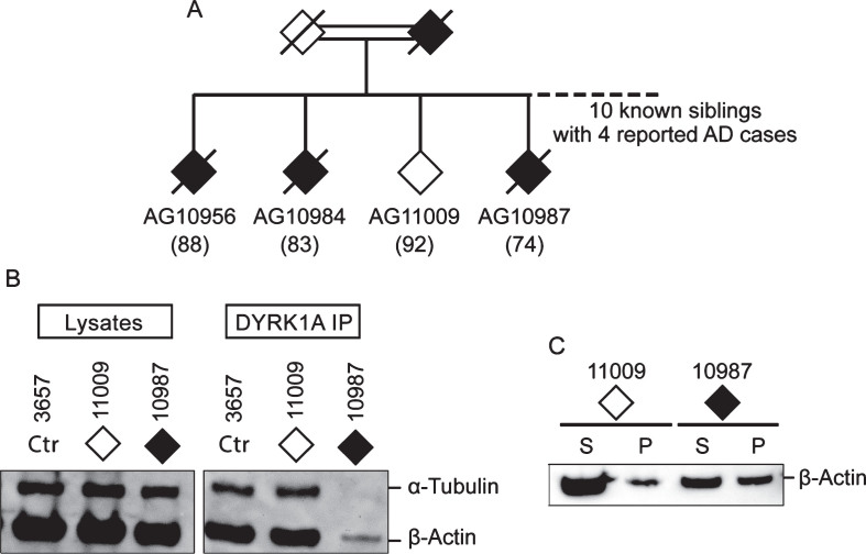
Our original observations of marked reduction of cytoskeletal proteins associated with DYRK1A in brains and lymphoblastoid cell lines from DS and AD prompted an investigation whether cytoskeleton abnormalities could potentially be used as biomarkers of AD.
Our assay relied on quantification of co-immunoprecipitated cytoskeletal proteins with DYRK1A (co-IP assay) and analysis of the profile of G- and F-actin fractions obtained by high-speed centrifugations (spin-down assay).
In co-IP assay, both DS and AD samples displayed reduced abundance of associated cytoskeletal proteins. In spin-down assay, G-actin fractions of controls displayed two closely spaced bands of actin in SDS-PAGE; while in AD and DS, only the upper band of the doublet was present. In both assays, alterations of actin cytoskeleton were present in DS, sporadic and familial AD cases, and in asymptomatic persons who later progressed to confirmed AD, but not in non-AD donors. In blind testing involving six AD and six controls, the above tests positively identified ten cases. Analysis of blood samples revealed the diversity of mild cognitive impairment (MCI) cases regarding the presence of the AD biomarker allowing distinction between likely prodromal AD and non-AD MCI cases.
Both brain tissue and lymphocytes from DS and AD displayed similar semi-quantitative and qualitative alterations of actin cytoskeleton. Their specificity for AD-type dementia and the presence before clinical onset of the disease make them suitable biomarker candidates for early and definite diagnosis of AD. The presence of alterations in peripheral tissue points to systemic underlying mechanisms and suggests that early dysfunction of cytoskeleton may be a predisposing factor in the development of AD.
双重特异性酪氨酸磷酸化调节激酶1A(DYRK1A)与智力迟钝以及唐氏综合征(DS)个体的阿尔茨海默病(AD)痴呆有关。该蛋白与细胞骨架相关,并且已表明表达改变会通过剂量效应损害细胞骨架网络。
我们最初观察到DS和AD患者大脑及淋巴母细胞系中与DYRK1A相关的细胞骨架蛋白显著减少,这促使我们研究细胞骨架异常是否有可能用作AD的生物标志物。
我们的检测方法依赖于对与DYRK1A共免疫沉淀的细胞骨架蛋白进行定量(共免疫沉淀检测),以及对高速离心获得的G-肌动蛋白和F-肌动蛋白组分的分布进行分析(沉降检测)。
在共免疫沉淀检测中,DS和AD样本中相关细胞骨架蛋白的丰度均降低。在沉降检测中,对照组的G-肌动蛋白组分在十二烷基硫酸钠-聚丙烯酰胺凝胶电泳(SDS-PAGE)中显示出两条紧密相邻的肌动蛋白条带;而在AD和DS样本中,仅出现双峰中的上条带。在这两种检测中,DS、散发性和家族性AD病例以及后来发展为确诊AD的无症状个体中均存在肌动蛋白细胞骨架改变,但在非AD供体中未出现。在涉及6例AD患者和6例对照的盲法检测中,上述检测方法正确识别出10例病例。对血样的分析揭示了轻度认知障碍(MCI)病例在AD生物标志物存在方面的多样性,从而能够区分可能的前驱AD和非AD MCI病例。
DS和AD患者的脑组织和淋巴细胞均显示出肌动蛋白细胞骨架类似的半定量和定性改变。它们对AD型痴呆的特异性以及在疾病临床发作前的存在使其成为AD早期和明确诊断的合适生物标志物候选物。外周组织中存在改变表明存在系统性潜在机制,并提示细胞骨架的早期功能障碍可能是AD发生发展的一个易感因素。