Haenel Alexander, Ghosn Mohamad, Karimi Tahereh, Vykoukal Jody, Shah Dipan, Valderrabano Miguel, Schulz Daryl G, Raizner Albert, Schmitz Christoph, Alt Eckhard U
Heart and Vascular Institute, Department of Medicine, Tulane University Health Science Center, New Orleans, LA 70112, United States.
Houston Methodist DeBakey Heart and Vascular Center, Houston, TX 77030, United States.
World J Stem Cells. 2019 Oct 26;11(10):831-858. doi: 10.4252/wjsc.v11.i10.831.
Numerous studies investigated cell-based therapies for myocardial infarction (MI). The conflicting results of these studies have established the need for developing innovative approaches for applying cell-based therapy for MI. Experimental studies on animal models demonstrated the potential of fresh, uncultured, unmodified, autologous adipose-derived regenerative cells (UA-ADRCs) for treating acute MI. In contrast, studies on the treatment of chronic MI (CMI; > 4 wk post-MI) with UA-ADRCs have not been published so far. Among several methods for delivering cells to the myocardium, retrograde delivery into a temporarily blocked coronary vein has recently been demonstrated as an effective option.
To test the hypothesis that in experimentally-induced chronic myocardial infarction (CMI; > 4 wk post-MI) in pigs, retrograde delivery of fresh, uncultured, unmodified, autologous adipose-derived regenerative cells (UA-ADRCs) into a temporarily blocked coronary vein improves cardiac function and structure.
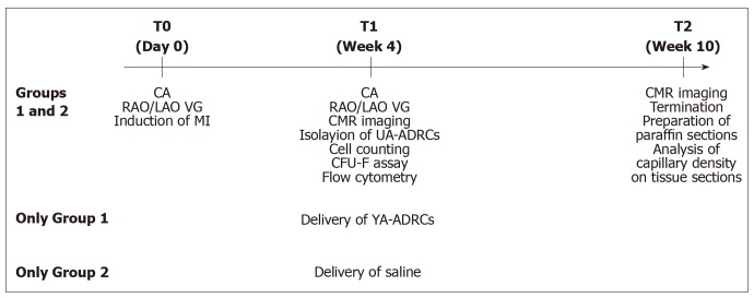
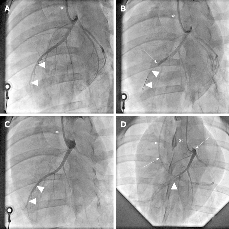
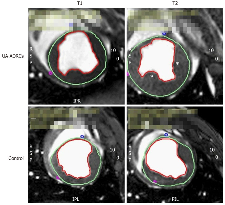
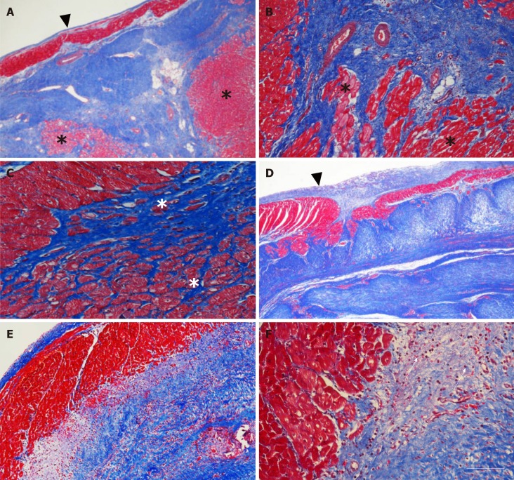
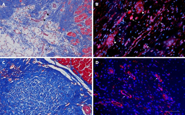
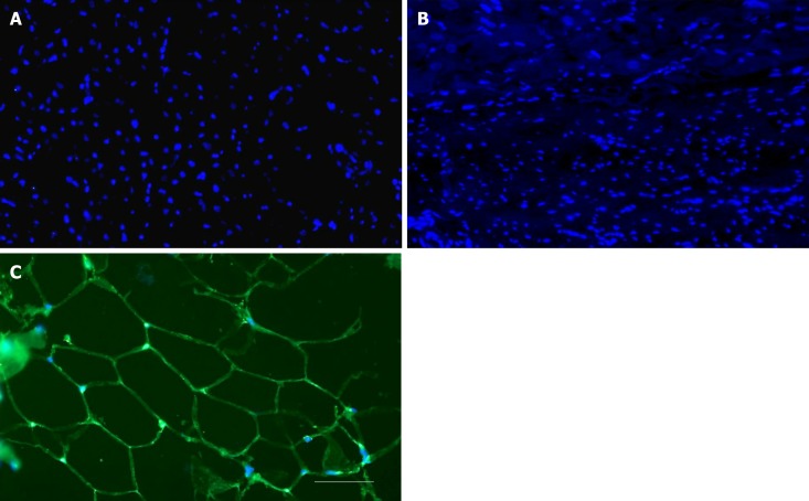
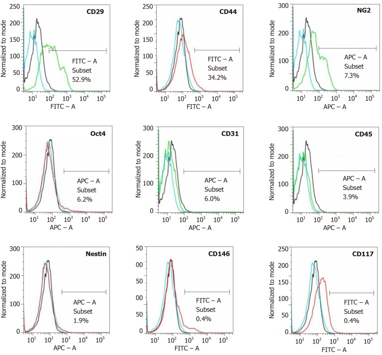
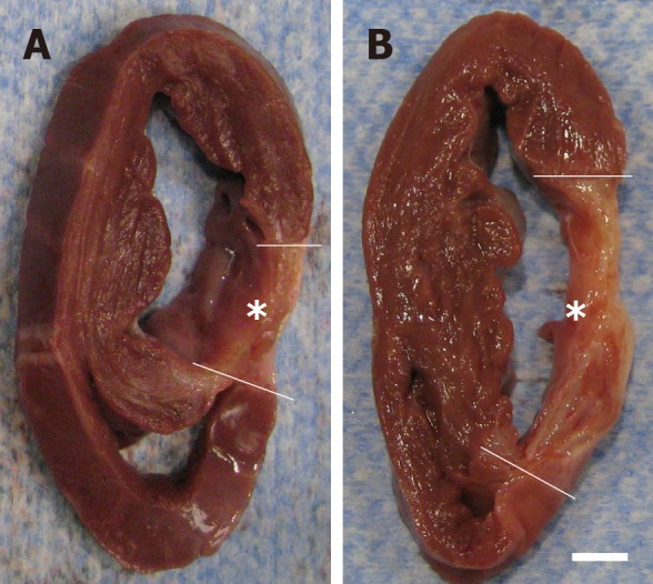
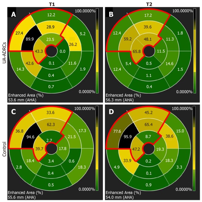
The left anterior descending (LAD) coronary artery of pigs was blocked for 180 min at time point T0. Then, either 18 × 10 UA-ADRCs prepared at "point of care" or saline as control were retrogradely delivered an over-the-wire balloon catheter placed in the temporarily blocked LAD vein 4 wk after T0 (T1). Effects of cells or saline were assessed by cardiac magnetic resonance (CMR) imaging, late gadolinium enhancement CMR imaging, and post mortem histologic analysis 10 wk after T0 (T2).
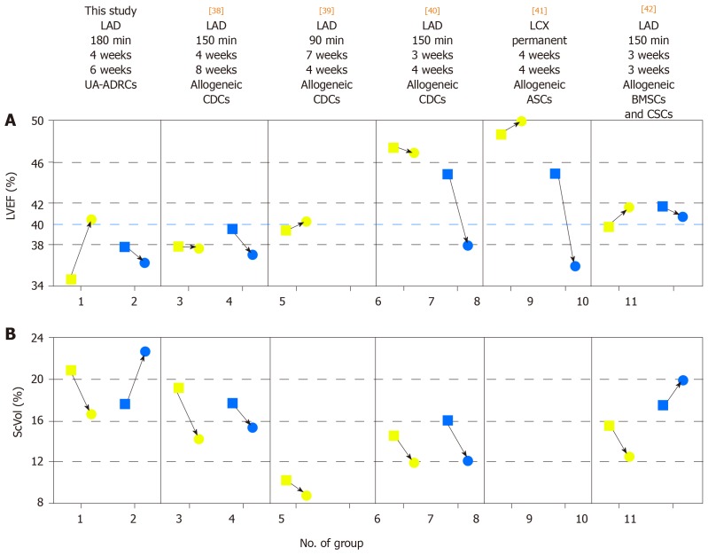
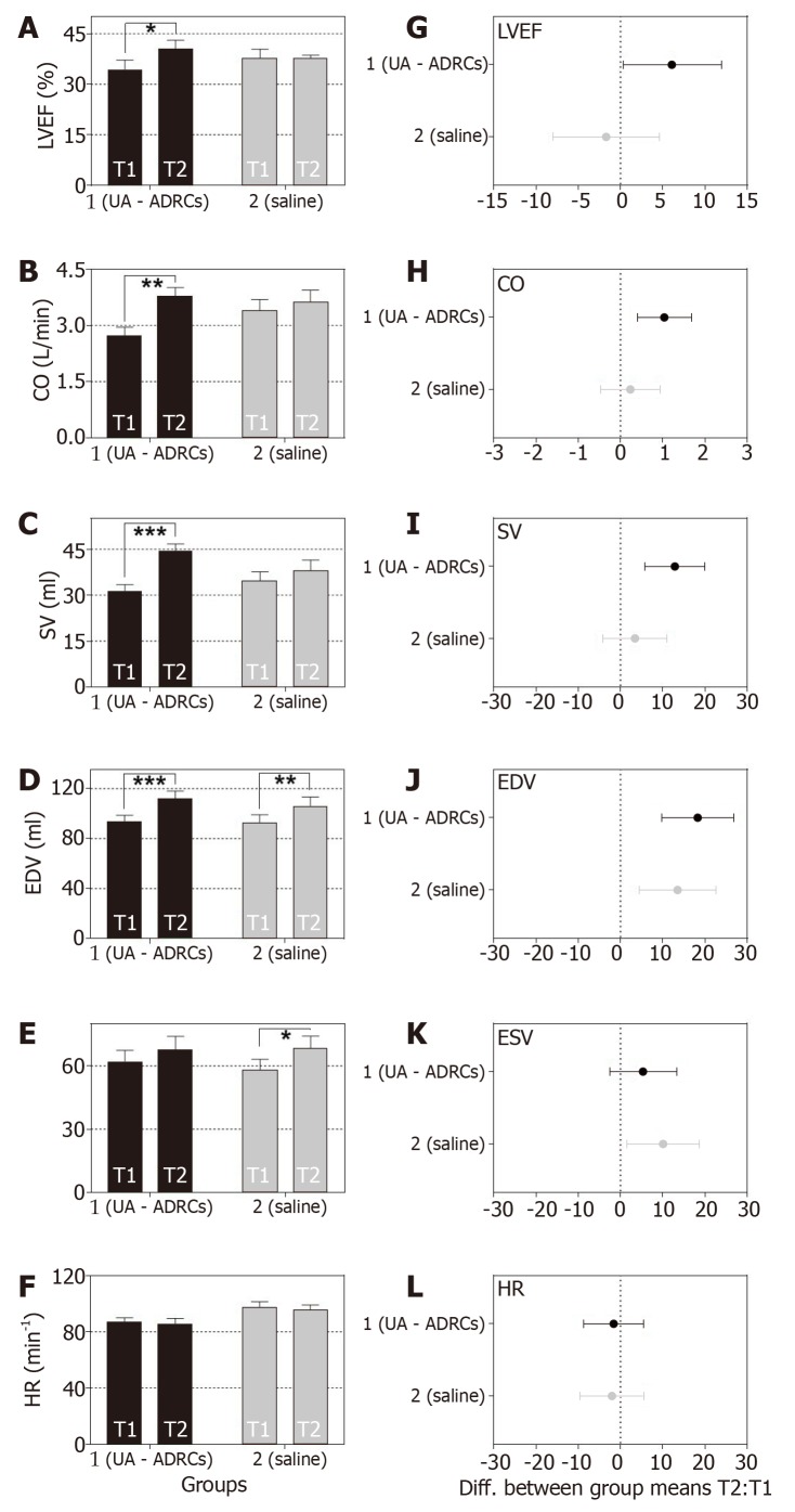
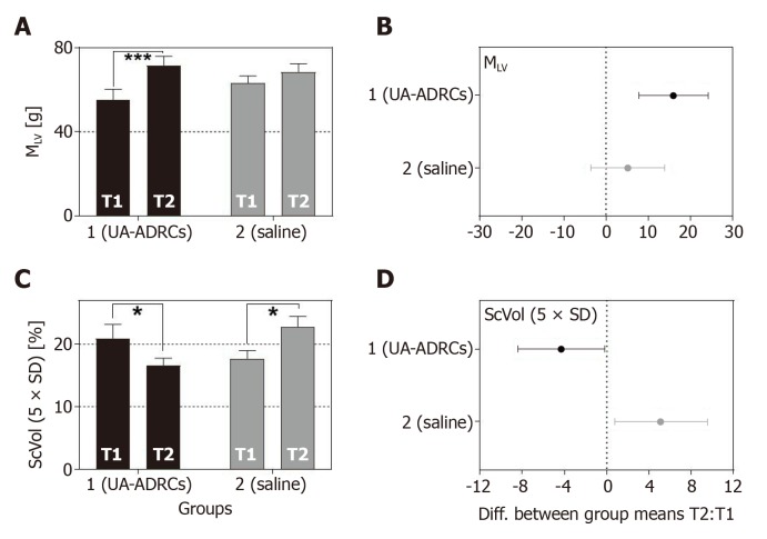
Unlike the delivery of saline, delivery of UA-ADRCs demonstrated statistically significant improvements in cardiac function and structure at T2 compared to T1 (all values given as mean ± SE): Increased mean LVEF (UA-ADRCs group: 34.3% ± 2.9% at T1 40.4 ± 2.6% at T2, = 0.037; saline group: 37.8% ± 2.6% at T1 36.2% ± 2.4% at T2, > 0.999), increased mean cardiac output (UA-ADRCs group: 2.7 ± 0.2 L/min at T1 3.8 ± 0.2 L/min at T2, = 0.002; saline group: 3.4 ± 0.3 L/min at T1 3.6 ± 0.3 L/min at T2, = 0.798), increased mean mass of the left ventricle (UA-ADRCs group: 55.3 ± 5.0 g at T1 71.3 ± 4.5 g at T2, < 0.001; saline group: 63.2 ± 3.4 g at T1 68.4 ± 4.0 g at T2, = 0.321) and reduced mean relative amount of scar volume of the left ventricular wall (UA-ADRCs group: 20.9% ± 2.3% at T1 16.6% ± 1.2% at T2, = 0.042; saline group: 17.6% ± 1.4% at T1 22.7% ± 1.8% at T2, = 0.022).
Retrograde cell delivery of UA-ADRCs in a porcine model for the study of CMI significantly improved myocardial function, increased myocardial mass and reduced the formation of scar tissue.
众多研究对心肌梗死(MI)的细胞疗法进行了调查。这些研究结果相互矛盾,因此有必要开发创新方法来应用细胞疗法治疗MI。在动物模型上进行的实验研究表明,新鲜、未培养、未修饰的自体脂肪源性再生细胞(UA-ADRCs)具有治疗急性MI的潜力。相比之下,目前尚未发表关于UA-ADRCs治疗慢性MI(CMI;MI后>4周)的研究。在几种将细胞递送至心肌的方法中,最近已证明经逆行将细胞递送至暂时阻断的冠状静脉是一种有效的选择。
验证以下假设:在实验诱导的猪慢性心肌梗死(CMI;MI后>4周)中,经逆行将新鲜、未培养、未修饰的自体脂肪源性再生细胞(UA-ADRCs)递送至暂时阻断的冠状静脉可改善心脏功能和结构。
在时间点T0将猪的左前降支(LAD)冠状动脉阻断180分钟。然后,在T0后4周(T1),将在“床边”制备的18×10个UA-ADRCs或作为对照的生理盐水经置于暂时阻断的LAD静脉中的过线球囊导管逆行递送。在T0后10周(T2),通过心脏磁共振(CMR)成像、钆增强延迟CMR成像和死后组织学分析评估细胞或生理盐水的作用。
与递送生理盐水不同,与T1相比,在T2时,UA-ADRCs的递送在心脏功能和结构方面显示出统计学上的显著改善(所有值均以平均值±标准误给出):左心室射血分数(LVEF)平均值增加(UA-ADRCs组:T1时为34.3%±
2.9%,T2时为40.4±2.6%,P = 0.037;生理盐水组:T1时为37.8%±2.6%,T2时为36.2%±2.4%,P>0.999),心输出量平均值增加(UA-ADRCs组:T1时为2.7±0.2 L/min,T2时为3.8±0.2 L/min,P = 0.002;生理盐水组:T1时为3.4±0.3 L/min,T2时为3.6±0.3 L/min,P = 0.798),左心室平均质量增加(UA-ADRCs组:T1时为55.3±5.0 g,T2时为71.3±4.5 g,P<0.001;生理盐水组:T1时为63.2±3.4 g,T2时为68.4±4.0 g,P = 0.321),左心室壁瘢痕体积的平均相对量减少(UA-ADRCs组:T1时为20.9%±2.3%,T2时为16.6%±1.2%,P = 0.042;生理盐水组:T1时为17.6%±1.4%,T2时为22.7%±1.8%,P = 0.022)。
在用于研究CMI的猪模型中,经逆行递送UA-ADRCs可显著改善心肌功能,增加心肌质量并减少瘢痕组织的形成。