Institute of Neuroscience, Newcastle University, Newcastle upon Tyne, UK.
Population Health and Immunity Division, The Walter and Eliza Hall Institute of Medical Research, Parkville, VIC, Australia.
Aust N Z J Psychiatry. 2020 Jun;54(6):633-643. doi: 10.1177/0004867419885165. Epub 2019 Nov 7.
We investigated the structural changes associated with Alzheimer's disease, dementia with Lewy bodies and Parkinson disease dementia by means of cortical thickness analysis.
Two hundred and forty-five participants: 76 Alzheimer's disease, 65 dementia with Lewy bodies, 29 Parkinson disease dementia and 76 cognitively normal controls underwent 3-T T1-weighted magnetic resonance imaging and clinical and cognitive assessments. We implemented FreeSurfer to obtain cortical thickness estimates to contrast patterns of cortical thinning across groups and their clinical correlates.
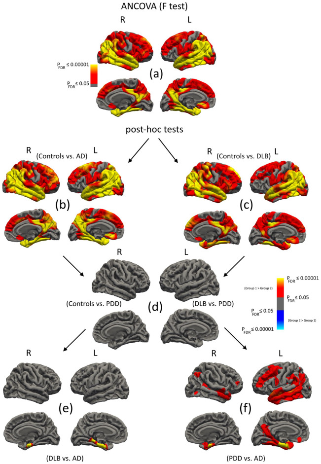
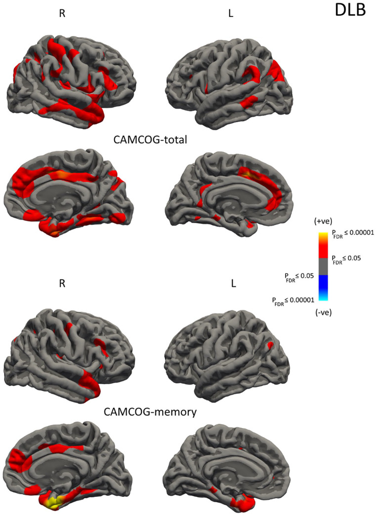
In Alzheimer's disease and dementia with Lewy bodies, a largely similar pattern of regional cortical thinning was observed relative to controls apart from a more severe loss within the entorhinal and parahippocampal structures in Alzheimer's disease. In Parkinson disease dementia, regional cortical thickness was indistinguishable from controls and dementia with Lewy bodies, suggesting an 'intermediate' pattern of regional cortical change. In terms of global cortical thickness, group profiles were controls > Parkinson disease dementia > dementia with Lewy bodies > Alzheimer's disease (F ⩽ 123.2, < 0.001), where percentage wise, the average difference compared to controls were -1.8%, -5.5% and -6.4%, respectively. In these samples, cortical thinning was also associated with cognitive decline in dementia with Lewy bodies but not in Parkinson disease dementia and Alzheimer's disease.
In a large and well-characterised cohort of people with dementia, regional cortical thinning in dementia with Lewy bodies was broadly similar to Alzheimer's disease. There was preservation of the medial temporal lobe structures in dementia with Lewy bodies compared with Alzheimer's disease, supporting its inclusion as a supportive biomarker in the revised clinical criteria for dementia with Lewy bodies. However, there was less global cortical thinning in Parkinson disease dementia, with no significant regional difference between Parkinson disease dementia and controls. These findings highlight the overlap across the Alzheimer's disease/Parkinson disease dementia spectrum and the potential for differing mechanisms underlying neurodegeneration and cognition in dementia with Lewy bodies and Parkinson disease dementia.
我们通过皮质厚度分析研究了与阿尔茨海默病、路易体痴呆和帕金森病痴呆相关的结构变化。
245 名参与者:76 名阿尔茨海默病患者,65 名路易体痴呆患者,29 名帕金森病痴呆患者和 76 名认知正常对照者接受了 3-T T1 加权磁共振成像和临床及认知评估。我们使用 FreeSurfer 获得皮质厚度估计值,以对比各组皮质变薄的模式及其与临床的相关性。
与对照组相比,阿尔茨海默病和路易体痴呆患者除了在阿尔茨海默病中出现更严重的内嗅皮质和海马旁回结构丢失外,还观察到大致相似的区域性皮质变薄模式。在帕金森病痴呆患者中,皮质厚度与对照组和路易体痴呆患者无明显差异,提示区域性皮质改变呈“中间”模式。在整体皮质厚度方面,各组的特征为对照组>帕金森病痴呆>路易体痴呆>阿尔茨海默病(F ⩽ 123.2,P < 0.001),与对照组相比,平均差异分别为-1.8%、-5.5%和-6.4%。在这些样本中,皮质变薄也与路易体痴呆患者的认知能力下降有关,但与帕金森病痴呆和阿尔茨海默病患者无关。
在一个由大量特征明确的痴呆患者组成的队列中,路易体痴呆患者的区域性皮质变薄与阿尔茨海默病患者大致相似。与阿尔茨海默病相比,路易体痴呆患者的内侧颞叶结构得以保留,支持将其作为路易体痴呆修订临床标准的支持性生物标志物。然而,帕金森病痴呆患者的皮质整体变薄程度较低,帕金森病痴呆患者与对照组之间无明显的皮质区域差异。这些发现突显了阿尔茨海默病/帕金森病痴呆谱系中的重叠,以及路易体痴呆和帕金森病痴呆中神经退行性变和认知的潜在不同机制。