Laboratorio de Procesado de Imagen (LPI), Universidad de Valladolid, 47011, Valladolid, Spain.
Cardiff University Brain Research Imaging Centre (CUBRIC), Cardiff University, Cardiff, CF24 4HQ, UK.
J Neurol. 2023 Jan;270(1):13-31. doi: 10.1007/s00415-022-11398-z. Epub 2022 Sep 30.
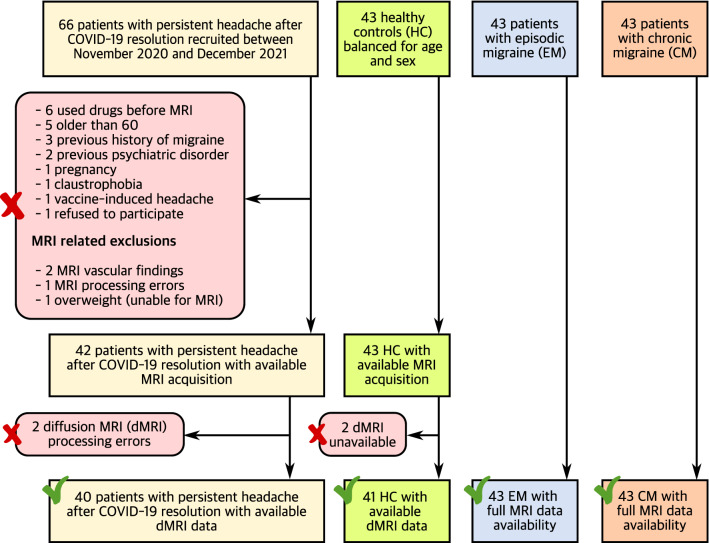
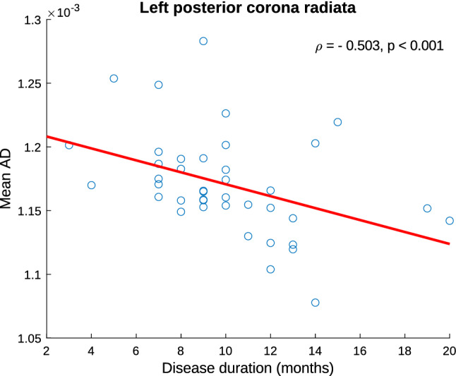
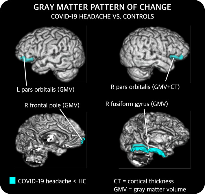
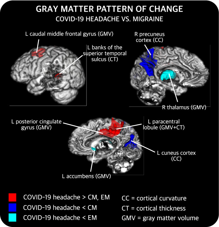
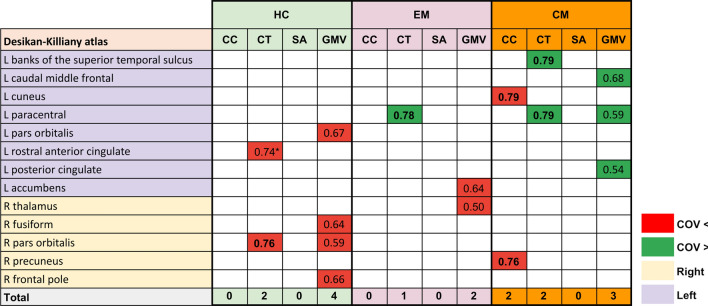
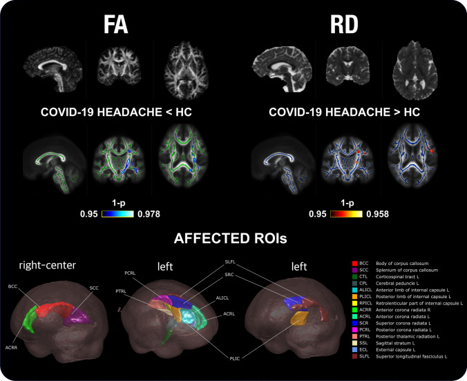
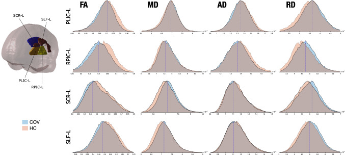
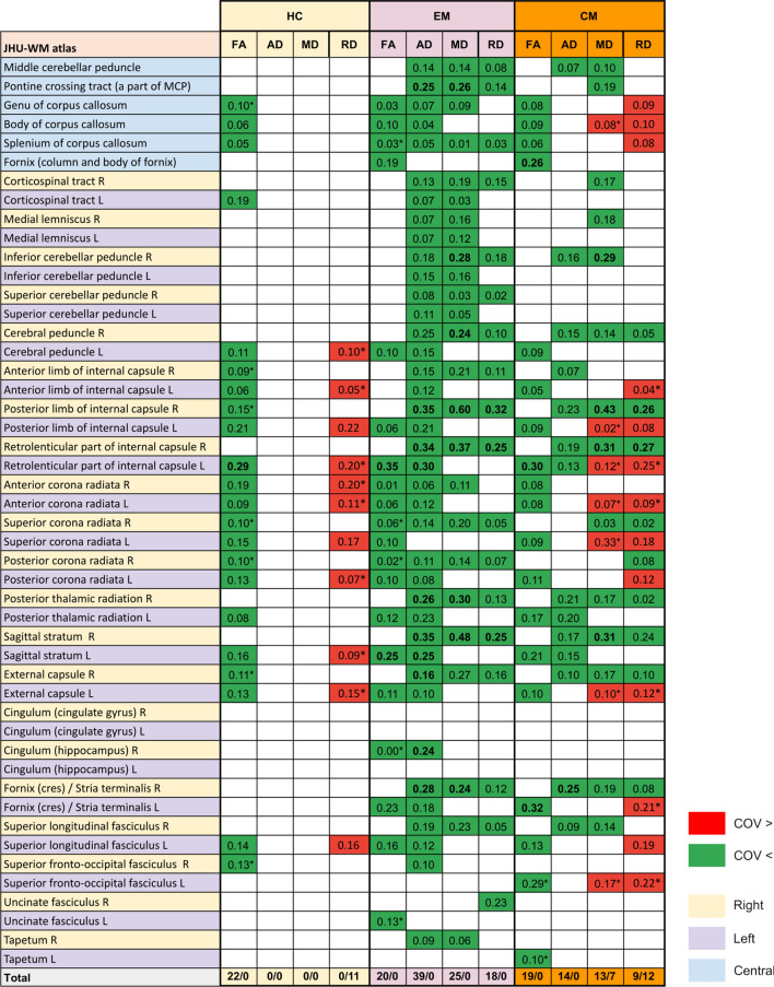
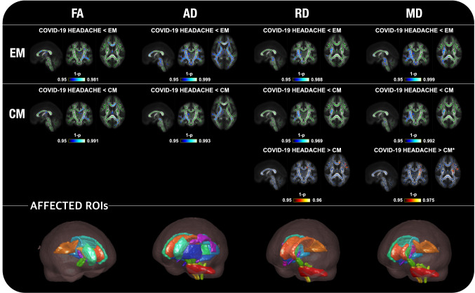
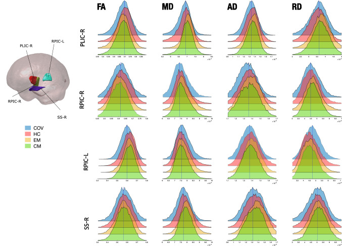
Headache is among the most frequently reported symptoms after resolution of COVID-19. We assessed structural brain changes using T1- and diffusion-weighted MRI processed data from 167 subjects: 40 patients who recovered from COVID-19 but suffered from persistent headache without prior history of headache (COV), 41 healthy controls, 43 patients with episodic migraine and 43 patients with chronic migraine. To evaluate gray matter and white matter changes, morphometry parameters and diffusion tensor imaging-based measures were employed, respectively. COV patients showed significant lower cortical gray matter volume and cortical thickness than healthy subjects (p < 0.05, false discovery rate corrected) in the inferior frontal and the fusiform cortex. Lower fractional anisotropy and higher radial diffusivity (p < 0.05, family-wise error corrected) were observed in COV patients compared to controls, mainly in the corpus callosum and left hemisphere. COV patients showed higher cortical volume and thickness than migraine patients in the cingulate and frontal gyri, paracentral lobule and superior temporal sulcus, lower volume in subcortical regions and lower curvature in the precuneus and cuneus. Lower diffusion metric values in COV patients compared to migraine were identified prominently in the right hemisphere. COV patients present diverse changes in the white matter and gray matter structure. White matter changes seem to be associated with impairment of fiber bundles. Besides, the gray matter changes and other white matter modifications such as axonal integrity loss seemed subtle and less pronounced than those detected in migraine, showing that persistent headache after COVID-19 resolution could be an intermediate state between normality and migraine.
头痛是 COVID-19 痊愈后最常报告的症状之一。我们评估了 167 名受试者的 T1 加权和弥散加权 MRI 处理数据的结构脑变化:40 名从 COVID-19 中康复但患有持续性头痛且无既往头痛史的患者(COV),41 名健康对照者,43 名发作性偏头痛患者和 43 名慢性偏头痛患者。为了评估灰质和白质的变化,分别采用了形态计量学参数和弥散张量成像测量方法。与健康对照组相比,COV 患者的额下回和梭状回的皮质灰质体积和皮质厚度显著降低(p<0.05,经假发现率校正)。与对照组相比,COV 患者的各向异性分数(FA)降低和径向弥散度(RD)升高(p<0.05,经全方差校正),主要在胼胝体和左半球。与偏头痛患者相比,COV 患者的扣带回和额回、旁中央小叶和颞上沟的皮质体积和厚度更高,皮质下区域的体积更小,楔前叶和楔叶的曲率更小。与偏头痛患者相比,COV 患者的右侧大脑半球的弥散测量值变化更为明显。COV 患者的白质和灰质结构均有不同程度的改变。白质变化似乎与纤维束的损伤有关。此外,与偏头痛患者相比,COV 患者的灰质变化和其他白质改变,如轴突完整性丧失,似乎较为轻微且不那么明显,这表明 COVID-19 痊愈后持续性头痛可能是介于正常和偏头痛之间的一种中间状态。