• 文献检索
  • 文档翻译
  • 深度研究
  • 学术资讯
  • Suppr Zotero 插件Zotero 插件
  • 邀请有礼
  • 套餐&价格
  • 历史记录
应用&插件
Suppr Zotero 插件Zotero 插件浏览器插件Mac 客户端Windows 客户端微信小程序
定价
高级版会员购买积分包购买API积分包
服务
文献检索文档翻译深度研究API 文档MCP 服务
关于我们
关于 Suppr公司介绍联系我们用户协议隐私条款
关注我们

Suppr 超能文献

核心技术专利:CN118964589B侵权必究
粤ICP备2023148730 号-1Suppr @ 2026

文献检索

告别复杂PubMed语法,用中文像聊天一样搜索,搜遍4000万医学文献。AI智能推荐,让科研检索更轻松。

立即免费搜索

文件翻译

保留排版,准确专业,支持PDF/Word/PPT等文件格式,支持 12+语言互译。

免费翻译文档

深度研究

AI帮你快速写综述,25分钟生成高质量综述,智能提取关键信息,辅助科研写作。

立即免费体验

仰卧位、俯卧位、右侧卧位和左侧卧位对人体肺循环的影响。

Supine, prone, right and left gravitational effects on human pulmonary circulation.

机构信息

Department of Clinical Physiology, Karolinska Institute, and Karolinska University Hospital, Stockholm, Sweden.

Department of Physiology and Pharmacology, Section of Anesthesiology and Intensive Care Medicine, Karolinska Institutet, and Function Perioperative Medicine and Intensive Care, Karolinska University Hospital, Stockholm, Sweden.

出版信息

J Cardiovasc Magn Reson. 2019 Nov 11;21(1):69. doi: 10.1186/s12968-019-0577-9.

DOI:10.1186/s12968-019-0577-9
PMID:31707989
原文链接:https://pmc.ncbi.nlm.nih.gov/articles/PMC6844066/
Abstract

BACKGROUND

Body position can be optimized for pulmonary ventilation/perfusion matching during surgery and intensive care. However, positional effects upon distribution of pulmonary blood flow and vascular distensibility measured as the pulmonary blood volume variation have not been quantitatively characterized. In order to explore the potential clinical utility of body position as a modulator of pulmonary hemodynamics, we aimed to characterize gravitational effects upon distribution of pulmonary blood flow, pulmonary vascular distension, and pulmonary vascular distensibility.

METHODS

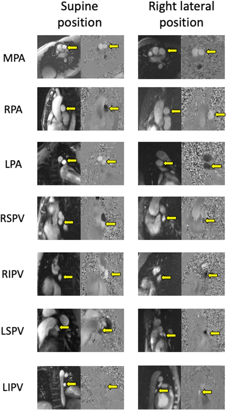
Healthy subjects (n = 10) underwent phase contrast cardiovascular magnetic resonance (CMR) pulmonary artery and vein flow measurements in the supine, prone, and right/left lateral decubitus positions. For each lung, blood volume variation was calculated by subtracting venous from arterial flow per time frame.

RESULTS

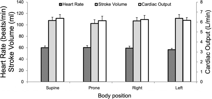
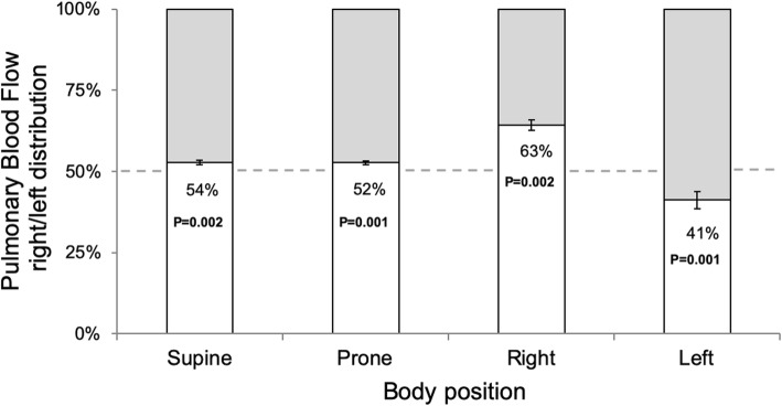
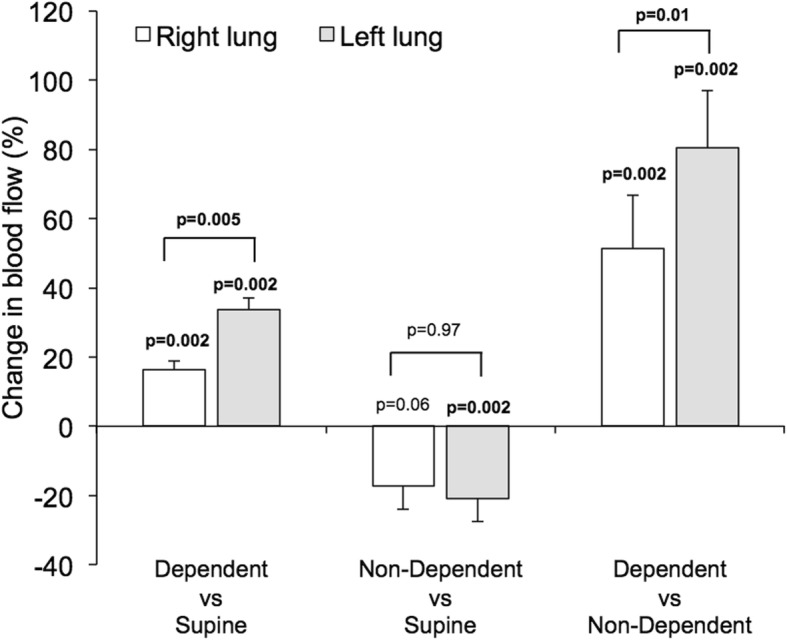
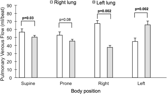
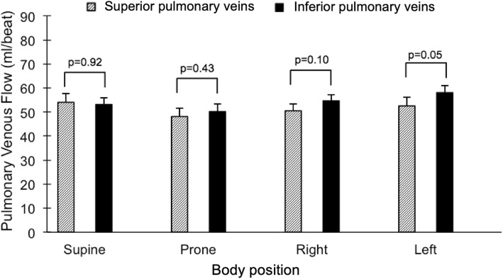
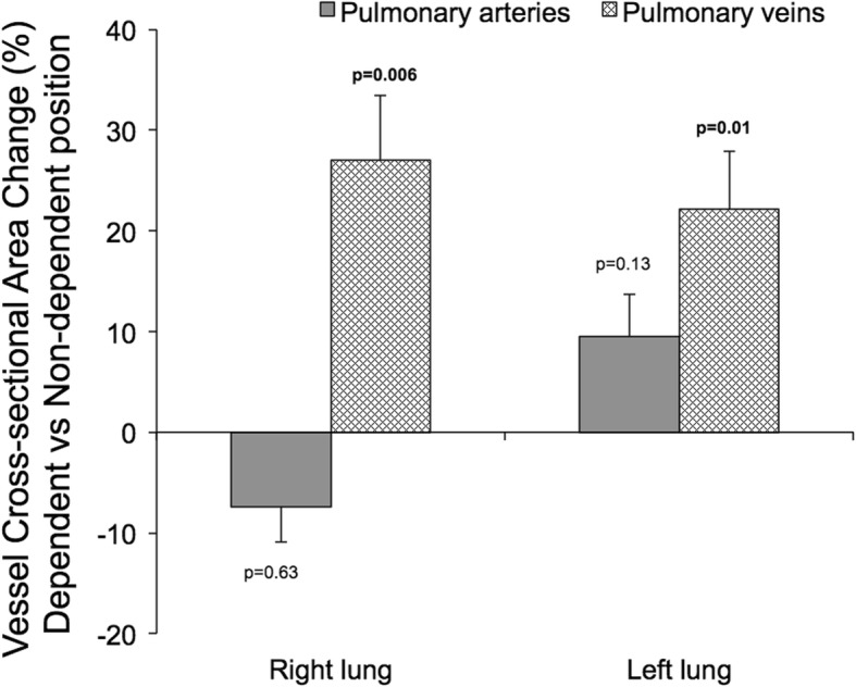
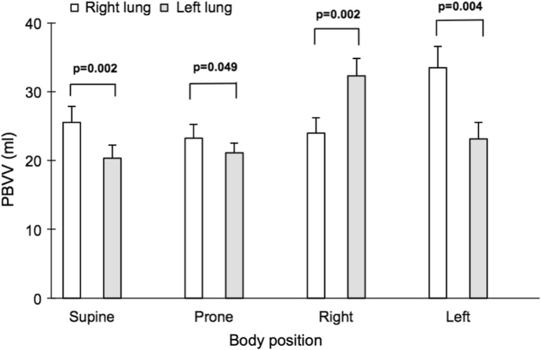
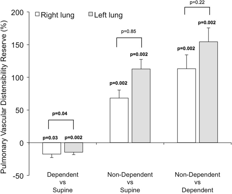
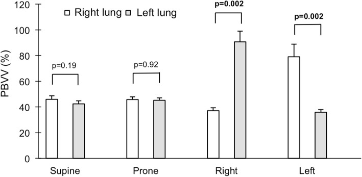
Body position did not change cardiac output (p = 0.84). There was no difference in blood flow between the superior and inferior pulmonary veins in the supine (p = 0.92) or prone body positions (p = 0.43). Compared to supine, pulmonary blood flow increased to the dependent lung in the lateral positions (16-33%, p = 0.002 for both). Venous but not arterial cross-sectional vessel area increased in both lungs when dependent compared to when non-dependent in the lateral positions (22-27%, p ≤ 0.01 for both). In contrast, compared to supine, distensibility increased in the non-dependent lung in the lateral positions (68-113%, p = 0.002 for both).

CONCLUSIONS

CMR demonstrates that in the lateral position, there is a shift in blood flow distribution, and venous but not arterial blood volume, from the non-dependent to the dependent lung. The non-dependent lung has a sizable pulmonary vascular distensibility reserve, possibly related to left atrial pressure. These results support the physiological basis for positioning patients with unilateral pulmonary pathology with the "good lung down" in the context of intensive care. Future studies are warranted to evaluate the diagnostic potential of these physiological insights into pulmonary hemodynamics, particularly in the context of non-invasively characterizing pulmonary hypertension.

摘要

背景

在手术和重症监护期间,可以优化体位以实现肺通气/灌注匹配。然而,尚未对体位对肺血流量分布和血管扩张性(以肺血容量变化来衡量)的影响进行定量描述。为了探索体位作为肺血流动力学调节剂的潜在临床应用,我们旨在描述体位对肺血流量分布、肺血管扩张和肺血管扩张性的影响。

方法

10 名健康受试者接受了仰卧位、俯卧位和右侧/左侧侧卧位下的相位对比心血管磁共振(CMR)肺动脉和静脉血流测量。对于每侧肺,通过从每个时间帧的动脉血流中减去静脉血流来计算血容量变化。

结果

体位不改变心输出量(p=0.84)。仰卧位和俯卧位时,上肺静脉和下肺静脉之间的血流没有差异(p=0.92 和 p=0.43)。与仰卧位相比,侧卧时肺血流量增加到下垂肺(16-33%,p=0.002 均)。与非下垂相比,侧卧时两侧肺的静脉而非动脉的血管横截面积在下垂时增加(22-27%,p=0.01 均)。相比之下,与仰卧位相比,侧卧时非下垂肺的顺应性增加(68-113%,p=0.002 均)。

结论

CMR 显示,在侧卧位时,血液从非下垂肺流向下垂肺,导致血流分布发生变化,静脉而非动脉的血容量发生变化。非下垂肺具有相当大的肺血管扩张储备能力,可能与左心房压力有关。这些结果支持在重症监护中对单侧肺病理患者采用“好肺在下”体位的生理基础。未来的研究需要评估这些对肺血流动力学的生理见解在诊断方面的潜力,特别是在无创性描述肺动脉高压方面。

https://cdn.ncbi.nlm.nih.gov/pmc/blobs/9132/6844066/bbba6e59eb33/12968_2019_577_Fig14_HTML.jpg
https://cdn.ncbi.nlm.nih.gov/pmc/blobs/9132/6844066/14b953a9c030/12968_2019_577_Fig1_HTML.jpg
https://cdn.ncbi.nlm.nih.gov/pmc/blobs/9132/6844066/521958a875ec/12968_2019_577_Fig2_HTML.jpg
https://cdn.ncbi.nlm.nih.gov/pmc/blobs/9132/6844066/2675bb26a595/12968_2019_577_Fig3_HTML.jpg
https://cdn.ncbi.nlm.nih.gov/pmc/blobs/9132/6844066/af9fc19b0af1/12968_2019_577_Fig4_HTML.jpg
https://cdn.ncbi.nlm.nih.gov/pmc/blobs/9132/6844066/7f3585a37c8f/12968_2019_577_Fig5_HTML.jpg
https://cdn.ncbi.nlm.nih.gov/pmc/blobs/9132/6844066/e7bb3e6d6d4b/12968_2019_577_Fig6_HTML.jpg
https://cdn.ncbi.nlm.nih.gov/pmc/blobs/9132/6844066/e763b9fb709d/12968_2019_577_Fig7_HTML.jpg
https://cdn.ncbi.nlm.nih.gov/pmc/blobs/9132/6844066/f529ec88442d/12968_2019_577_Fig8_HTML.jpg
https://cdn.ncbi.nlm.nih.gov/pmc/blobs/9132/6844066/75c18e22d5ee/12968_2019_577_Fig9_HTML.jpg
https://cdn.ncbi.nlm.nih.gov/pmc/blobs/9132/6844066/77ea28d11f08/12968_2019_577_Fig10_HTML.jpg
https://cdn.ncbi.nlm.nih.gov/pmc/blobs/9132/6844066/0f6db064d3a4/12968_2019_577_Fig11_HTML.jpg
https://cdn.ncbi.nlm.nih.gov/pmc/blobs/9132/6844066/f97bc936a98a/12968_2019_577_Fig12_HTML.jpg
https://cdn.ncbi.nlm.nih.gov/pmc/blobs/9132/6844066/038ea8c8f2fd/12968_2019_577_Fig13_HTML.jpg
https://cdn.ncbi.nlm.nih.gov/pmc/blobs/9132/6844066/bbba6e59eb33/12968_2019_577_Fig14_HTML.jpg

相似文献

1
Supine, prone, right and left gravitational effects on human pulmonary circulation.仰卧位、俯卧位、右侧卧位和左侧卧位对人体肺循环的影响。
J Cardiovasc Magn Reson. 2019 Nov 11;21(1):69. doi: 10.1186/s12968-019-0577-9.
2
Changes in systemic and pulmonary blood flow distribution in normal adult volunteers in response to posture and exercise: a phase contrast magnetic resonance imaging study.正常成年志愿者在姿势和运动反应下全身及肺血流分布的变化:一项相位对比磁共振成像研究。
J Physiol Sci. 2014 Mar;64(2):105-12. doi: 10.1007/s12576-013-0298-z. Epub 2014 Jan 3.
3
Pelvic cardiovascular magnetic resonance venography: venous changes with patient position and hydration status.盆腔心血管磁共振静脉造影:患者体位和水合状态对静脉变化的影响。
J Cardiovasc Magn Reson. 2019 Jan 3;21(1):3. doi: 10.1186/s12968-018-0503-6.
4
The gravitational distribution of ventilation-perfusion ratio is more uniform in prone than supine posture in the normal human lung.正常人体肺中,仰卧位时比俯卧位时通气-灌注比的重力分布更均匀。
J Appl Physiol (1985). 2013 Aug 1;115(3):313-24. doi: 10.1152/japplphysiol.01531.2012. Epub 2013 Apr 25.
5
Quantitative cardiovascular magnetic resonance in pregnant women: cross-sectional analysis of physiological parameters throughout pregnancy and the impact of the supine position.孕妇的定量心血管磁共振:整个孕期生理参数的横截面分析及仰卧位的影响。
J Cardiovasc Magn Reson. 2011 Jun 27;13(1):31. doi: 10.1186/1532-429X-13-31.
6
Pulmonary blood flow does not redistribute in dogs with reposition from supine to left lateral position.从仰卧位转为左侧卧位时,犬的肺血流不会重新分布。
Anesthesiology. 1998 Aug;89(2):483-92. doi: 10.1097/00000542-199808000-00026.
7
Influence of gravity on pulmonary venous flow velocity patterns: analysis of left and right pulmonary venous flow velocities in left and right decubitus positions.重力对肺静脉血流速度模式的影响:左右卧位时左右肺静脉血流速度分析
Am Heart J. 1999 Mar;137(3):419-26. doi: 10.1016/s0002-8703(99)70486-2.
8
Optimizing human pulmonary perfusion measurement using an in silico model of arterial spin labeling magnetic resonance imaging.使用动脉自旋标记磁共振成像的计算机模拟模型优化人体肺灌注测量。
Physiol Rep. 2019 Jun;7(11):e14077. doi: 10.14814/phy2.14077.
9
Pulmonary intravascular blood volume changes through the cardiac cycle in healthy volunteers studied by cardiovascular magnetic resonance measurements of arterial and venous flow.通过心血管磁共振测量动脉和静脉血流,对健康志愿者在心动周期中肺血管内血容量变化进行了研究。
J Cardiovasc Magn Reson. 2009 Oct 30;11(1):42. doi: 10.1186/1532-429X-11-42.
10
The effect of supine positioning on maternal hemodynamics during late pregnancy.仰卧位对妊娠晚期孕妇血流动力学的影响。
J Matern Fetal Neonatal Med. 2019 Dec;32(23):3923-3930. doi: 10.1080/14767058.2018.1478958. Epub 2018 Jun 3.

引用本文的文献

1
': congenital pericardial defect with structural, electrophysiological, and haemodynamic alterations induced by postural changes-a case report.先天性心包缺损伴体位改变引起的结构、电生理和血流动力学改变——病例报告
Eur Heart J Case Rep. 2025 Apr 21;9(5):ytaf200. doi: 10.1093/ehjcr/ytaf200. eCollection 2025 May.
2
Tracheal intubation in the lateral position in emergency medicine: a narrative review and clinical protocol.急诊医学中侧卧位气管插管:一篇叙述性综述及临床方案
World J Emerg Med. 2025;16(2):103-112. doi: 10.5847/wjem.j.1920-8642.2025.034.
3
Effect of Neck Rotation With Flexion on Ultrasonographic Optic Nerve Sheath Diameter in Patients Undergoing Elective Craniotomy.

本文引用的文献

1
Lateral positioning for critically ill adult patients.危重症成年患者的侧卧位摆放
Cochrane Database Syst Rev. 2016 May 12;2016(5):CD007205. doi: 10.1002/14651858.CD007205.pub2.
2
MR phase-contrast imaging in pulmonary hypertension.肺动脉高压中的磁共振相位对比成像
Br J Radiol. 2016 Jul;89(1063):20150995. doi: 10.1259/bjr.20150995. Epub 2016 Apr 6.
3
Validation and Development of a New Automatic Algorithm for Time-Resolved Segmentation of the Left Ventricle in Magnetic Resonance Imaging.磁共振成像中左心室时间分辨分割新自动算法的验证与开发
择期开颅手术患者颈部屈曲旋转对超声视神经鞘直径的影响。
Cureus. 2024 Mar 7;16(3):e55760. doi: 10.7759/cureus.55760. eCollection 2024 Mar.
4
Do blood flow patterns in the left atriums differ between left upper lobectomy and other lobectomies? A computational study.左上叶切除术与其他肺叶切除术相比,左心房的血流模式是否存在差异?一项计算研究。
Front Cardiovasc Med. 2024 Jan 5;10:1305526. doi: 10.3389/fcvm.2023.1305526. eCollection 2023.
5
Position-Dependent Symptoms of Pneumothorax During Mechanical Ventilation: A Case Report.机械通气期间气胸的体位相关症状:一例报告
Cureus. 2023 Dec 20;15(12):e50820. doi: 10.7759/cureus.50820. eCollection 2023 Dec.
6
Relationship between pulmonary blood flow and volume following lung resection using dynamic perfusion digital radiography.使用动态灌注数字射线摄影术评估肺切除术后肺血流量与肺容积之间的关系
J Thorac Dis. 2023 Oct 31;15(10):5593-5604. doi: 10.21037/jtd-23-986. Epub 2023 Oct 8.
7
Effects of intermittent pneumatic compression on the recovery of cardiovascular parameters after repeated sprint exercise.间歇式气动压迫对重复冲刺运动后心血管参数恢复的影响。
Eur J Appl Physiol. 2024 Apr;124(4):1037-1048. doi: 10.1007/s00421-023-05333-x. Epub 2023 Oct 4.
8
Pulmonary vein flow split effects in patient-specific simulations of left atrial flow.左心房血流患者特定模拟中的肺静脉血流分离效应。
Comput Biol Med. 2023 Sep;163:107128. doi: 10.1016/j.compbiomed.2023.107128. Epub 2023 Jun 12.
9
A method for calculating left ventricular end-diastolic volume as an index of left ventricular preload from the pre-ejection period, ejection time, blood pressure, and stroke volume: a prospective, observational study.一种从射血前期、射血时间、血压和每搏输出量计算左心室舒张末期容积作为左心室前负荷指数的方法:一项前瞻性观察研究。
BMC Anesthesiol. 2023 Apr 28;23(1):143. doi: 10.1186/s12871-023-02103-2.
10
Simulating Multi-Scale Pulmonary Vascular Function by Coupling Computational Fluid Dynamics With an Anatomic Network Model.通过将计算流体动力学与解剖网络模型相结合来模拟多尺度肺血管功能
Front Netw Physiol. 2022 Apr 25;2:867551. doi: 10.3389/fnetp.2022.867551. eCollection 2022.
Biomed Res Int. 2015;2015:970357. doi: 10.1155/2015/970357. Epub 2015 Jun 21.
4
Blood flow vortices along the main pulmonary artery measured with MR imaging for diagnosis of pulmonary hypertension.磁共振成像测量主肺动脉血流涡旋诊断肺动脉高压
Radiology. 2015 Apr;275(1):71-9. doi: 10.1148/radiol.14140849. Epub 2014 Nov 5.
5
Quantitative analysis of regional lung ventilation and perfusion PET with (68)Ga-labelled tracers.使用(68)Ga标记示踪剂对局部肺通气和灌注PET进行定量分析。
Nucl Med Commun. 2014 May;35(5):501-10. doi: 10.1097/MNM.0000000000000084.
6
Standardized cardiovascular magnetic resonance (CMR) protocols 2013 update.2013 年标准化心血管磁共振(CMR)协议更新。
J Cardiovasc Magn Reson. 2013 Oct 8;15(1):91. doi: 10.1186/1532-429X-15-91.
7
Pulmonary blood volume indexed to lung volume is reduced in newly diagnosed systemic sclerosis compared to normals--a prospective clinical cardiovascular magnetic resonance study addressing pulmonary vascular changes.与正常人相比,新诊断的系统性硬化症患者的肺血容量与肺容积的比值降低——一项前瞻性临床心血管磁共振研究探讨了肺血管变化。
J Cardiovasc Magn Reson. 2013 Sep 25;15(1):86. doi: 10.1186/1532-429X-15-86.
8
Determinants of pulmonary blood flow distribution.肺血流分布的决定因素。
Compr Physiol. 2011 Jan;1(1):39-59. doi: 10.1002/cphy.c090002.
9
Prone positioning in severe acute respiratory distress syndrome.俯卧位通气治疗严重急性呼吸窘迫综合征。
N Engl J Med. 2013 Jun 6;368(23):2159-68. doi: 10.1056/NEJMoa1214103. Epub 2013 May 20.
10
Pulmonary blood volume variation decreases after myocardial infarction in pigs: a quantitative and noninvasive MR imaging measure of heart failure.猪心肌梗死后肺血容量变化减少:心力衰竭的定量和无创性磁共振成像测量方法。
Radiology. 2010 Aug;256(2):415-23. doi: 10.1148/radiol.10090292.