University of Houston, College of Optometry, Houston, TX, United States of America.
King Abdullah University of Science and Technology (KAUST), Core Labs, Thuwal, Saudi Arabia.
PLoS One. 2019 Nov 13;14(11):e0224434. doi: 10.1371/journal.pone.0224434. eCollection 2019.
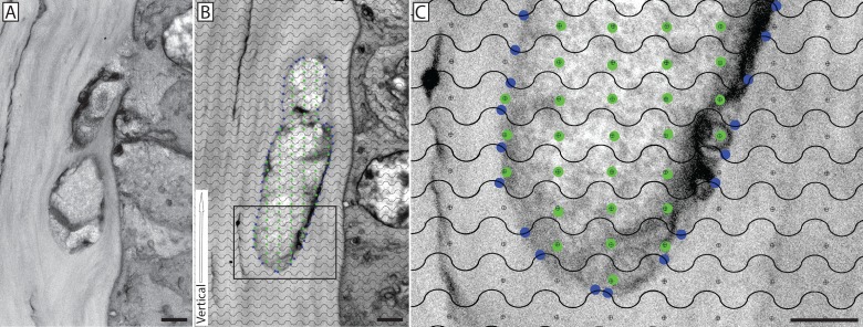
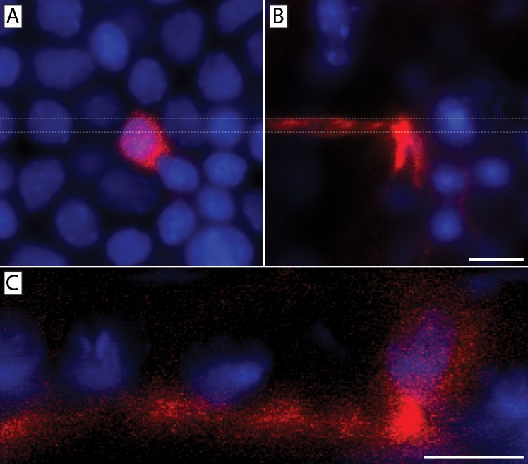
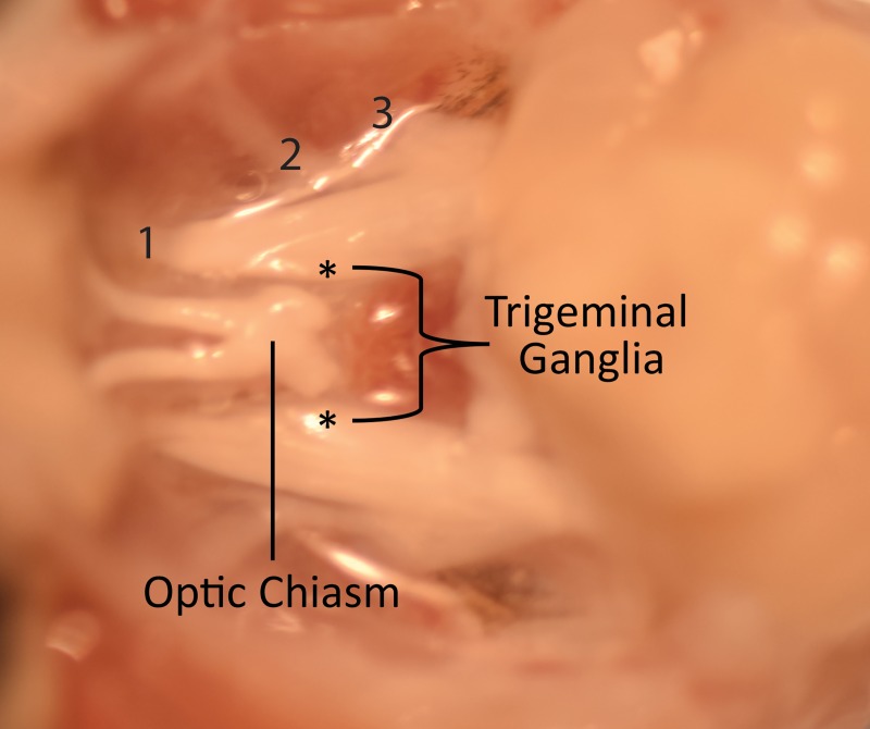
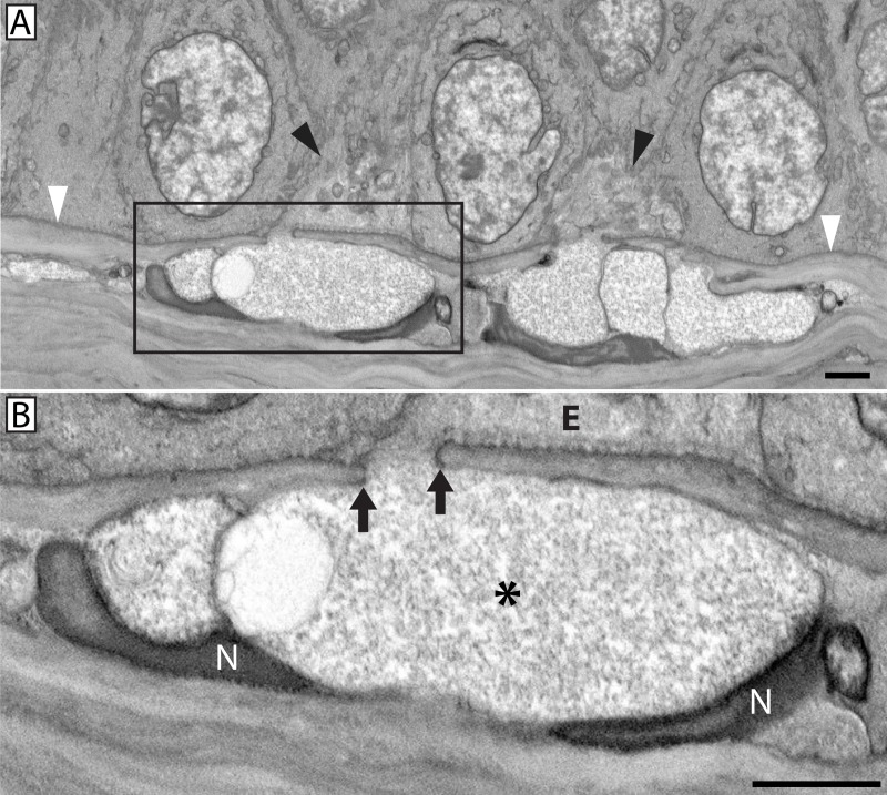
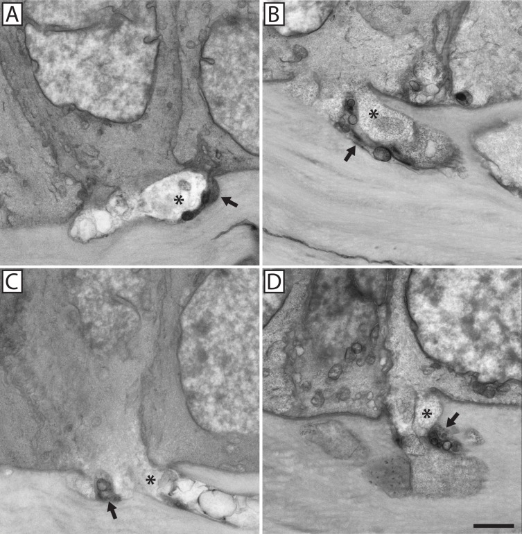
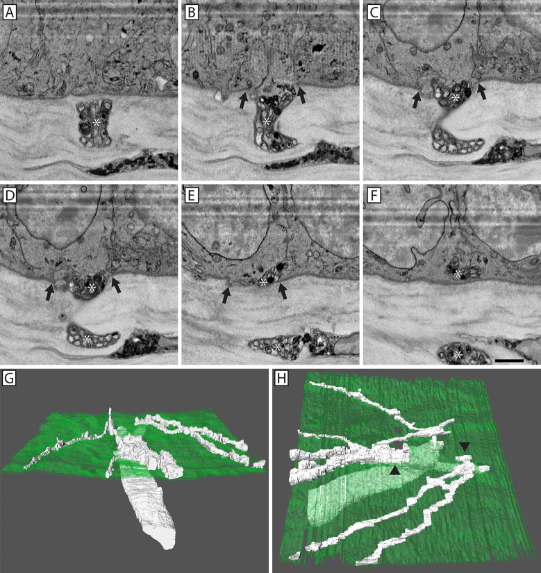
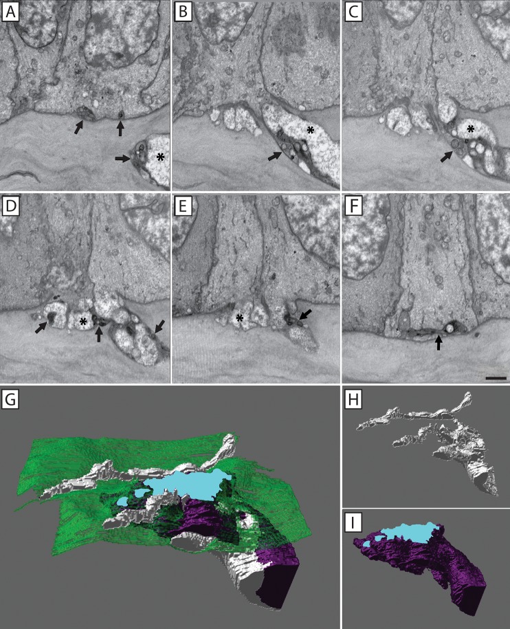
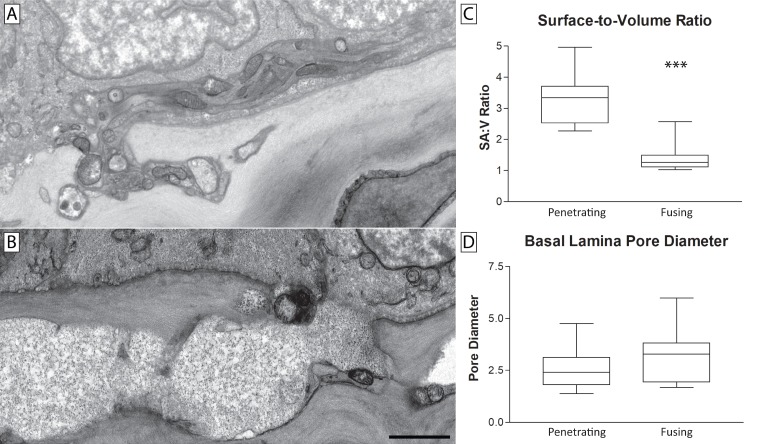
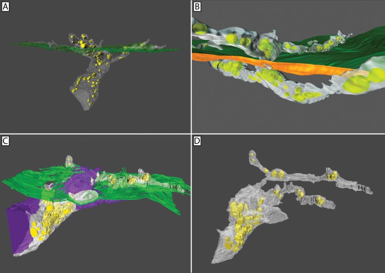
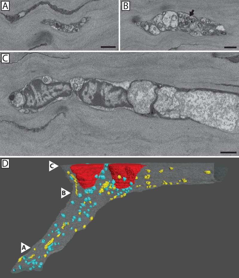
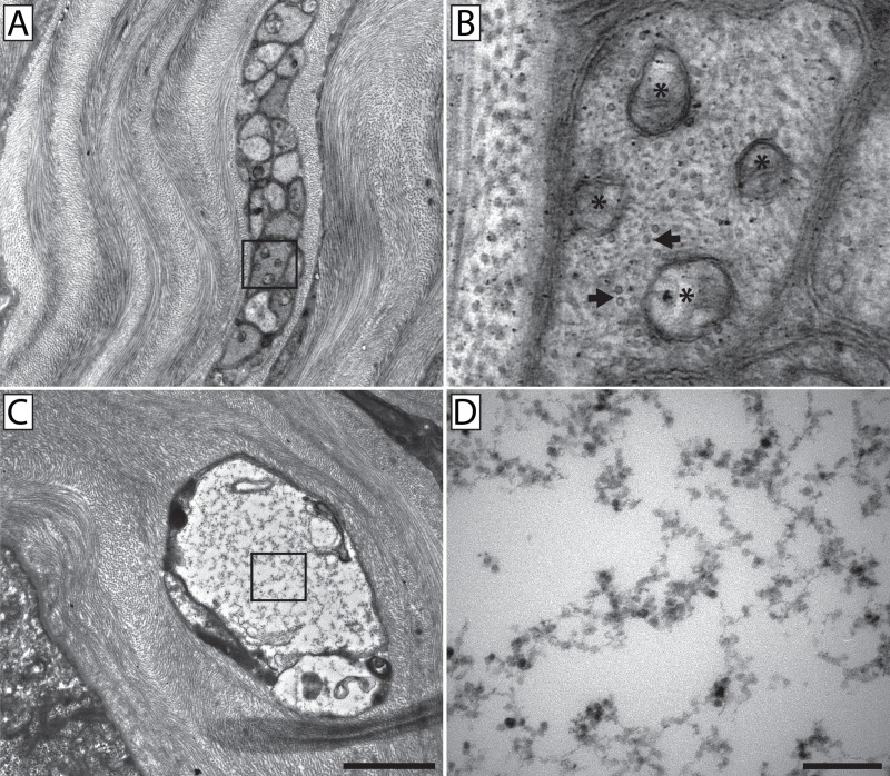
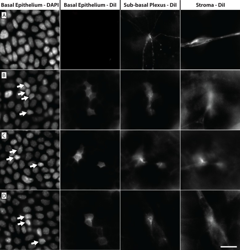
The cornea is the most highly innervated tissue in the body. It is generally accepted that corneal stromal nerves penetrate the epithelial basal lamina giving rise to intra-epithelial nerves. During the course of a study wherein we imaged corneal nerves in mice, we observed a novel neuronal-epithelial cell interaction whereby nerves approaching the epithelium in the cornea fused with basal epithelial cells, such that their plasma membranes were continuous and the neuronal axoplasm freely abutted the epithelial cytoplasm. In this study we sought to determine the frequency, distribution, and morphological profile of neuronal-epithelial cell fusion events within the cornea. Serial electron microscopy images were obtained from the anterior stroma in the paralimbus and central cornea of 8-10 week old C57BL/6J mice. We found evidence of a novel alternative behavior involving a neuronal-epithelial interaction whereby 42.8% of central corneal nerve bundles approaching the epithelium contain axons that fuse with basal epithelial cells. The average surface-to-volume ratio of a penetrating nerve was 3.32, while the average fusing nerve was smaller at 1.39 (p ≤ 0.0001). Despite this, both neuronal-epithelial cell interactions involve similarly sized discontinuities in the basal lamina. In order to verify the plasma membrane continuity between fused neurons and epithelial cells we used the lipophilic membrane tracer DiI. The majority of corneal nerves were labeled with DiI after application to the trigeminal ganglion and, consistent with our ultrastructural observations, fusion sites recognized as DiI-labeled basal epithelial cells were located at points of stromal nerve termination. These studies provide evidence that neuronal-epithelial cell fusion is a cell-cell interaction that occurs primarily in the central cornea, and fusing nerve bundles are morphologically distinct from penetrating nerve bundles. This is, to our knowledge, the first description of neuronal-epithelial cell fusion in the literature adding a new level of complexity to the current understanding of corneal innervation.
角膜是人体中神经分布最密集的组织。人们普遍认为,角膜基质神经穿透上皮基底层,形成上皮内神经。在一项研究中,我们对小鼠的角膜神经进行成像观察,发现了一种新的神经元-上皮细胞相互作用方式,即接近角膜上皮的神经与基底上皮细胞融合,使得它们的质膜连续,神经元轴突质自由毗邻上皮细胞质。在这项研究中,我们试图确定角膜内神经元-上皮细胞融合事件的频率、分布和形态特征。从 8-10 周龄 C57BL/6J 小鼠的前基质在旁角膜和中央角膜获得了连续的电子显微镜图像。我们发现了一种涉及神经元-上皮相互作用的新的替代行为的证据,即 42.8%的接近上皮的中央角膜神经束包含与基底上皮细胞融合的轴突。穿透神经的平均表面积与体积比为 3.32,而融合神经的平均表面积更小,为 1.39(p≤0.0001)。尽管如此,两种神经元-上皮细胞相互作用都涉及基底膜中类似大小的不连续性。为了验证融合神经元和上皮细胞之间的质膜连续性,我们使用了亲脂性膜示踪剂 DiI。将 DiI 应用于三叉神经节后,大多数角膜神经被标记,与我们的超微结构观察一致,融合部位被识别为 DiI 标记的基底上皮细胞,位于基质神经末端处。这些研究提供了证据表明,神经元-上皮细胞融合是一种细胞-细胞相互作用,主要发生在中央角膜,并且融合的神经束在形态上与穿透神经束不同。这是我们所知的文献中首次描述神经元-上皮细胞融合,为角膜神经支配的当前理解增加了一个新的复杂性水平。