Huda Fathul, Fan Yiping, Suzuki Mamiko, Konno Ayumu, Matsuzaki Yasunori, Takahashi Nobutaka, Chan Jerry K Y, Hirai Hirokazu
Department of Neurophysiology & Neural Repair, Gunma University Graduate School of Medicine, Maebashi, Gunma 371-8511, Japan.
Physiology Division, Department of Anatomy Physiology and Cell Biology, Faculty of Medicine Universitas Padjadjaran, Bandung, 40161, Indonesia.
PLoS One. 2016 Nov 1;11(11):e0164202. doi: 10.1371/journal.pone.0164202. eCollection 2016.
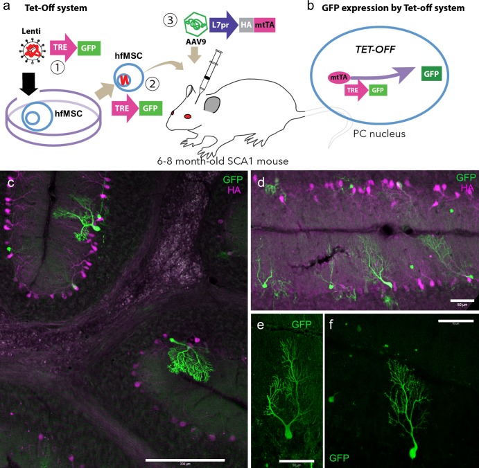
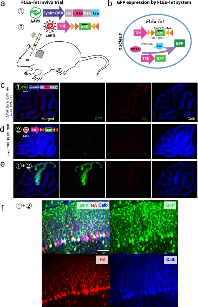
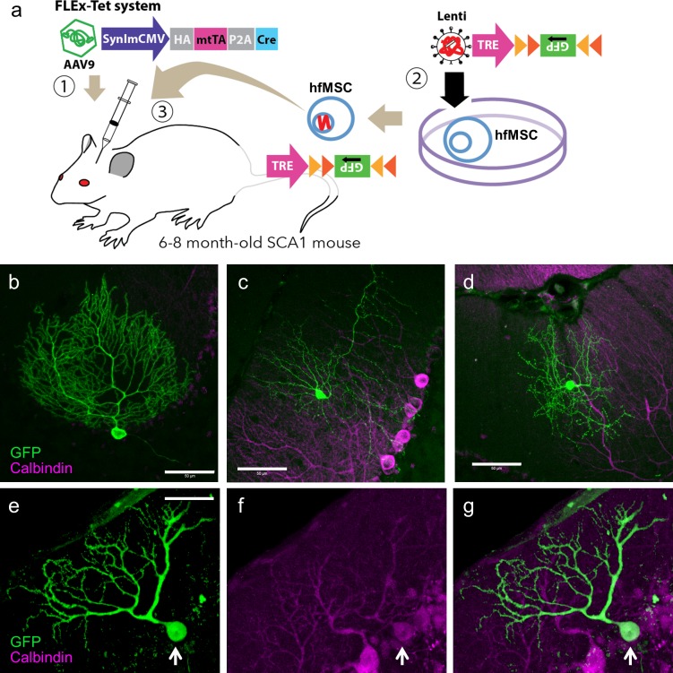
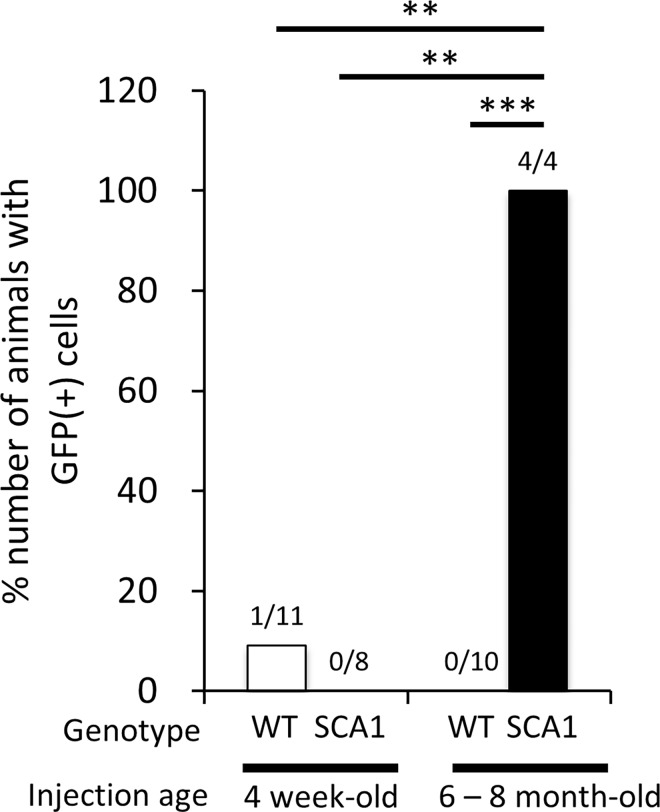
Mesenchymal stem cells (MSCs) migrate to damaged tissues, where they participate in tissue repair. Human fetal MSCs (hfMSCs), compared with adult MSCs, have higher proliferation rates, a greater differentiation capacity and longer telomeres with reduced senescence. Therefore, transplantation of quality controlled hfMSCs is a promising therapeutic intervention. Previous studies have shown that intravenous or intracortical injections of MSCs result in the emergence of binucleated cerebellar Purkinje cells (PCs) containing an MSC-derived marker protein in mice, thus suggesting a fusion event. However, transdifferentiation of MSCs into PCs or transfer of a marker protein from an MSC to a PC cannot be ruled out. In this study, we unequivocally demonstrated the fusion of hfMSCs with murine PCs through a tetracycline-regulated (Tet-off) system with or without a Cre-dependent genetic inversion switch (flip-excision; FLEx). In the FLEx-Tet system, we performed intra-cerebellar injection of viral vectors expressing tetracycline transactivator (tTA) and Cre recombinase into either non-symptomatic (4-week-old) or clearly symptomatic (6-8-month-old) spinocerebellar ataxia type 1 (SCA1) mice. Then, the mice received an injection of 50,000 genetically engineered hfMSCs that expressed GFP only in the presence of Cre recombinase and tTA. We observed a significant emergence of GFP-expressing PCs and interneurons in symptomatic, but not non-symptomatic, SCA1 mice 2 weeks after the MSC injection. These results, together with the results obtained using age-matched wild-type mice, led us to conclude that hfMSCs have the potential to preferentially fuse with degenerating PCs and interneurons but not with healthy neurons.
间充质干细胞(MSCs)迁移至受损组织,在那里参与组织修复。与成人间充质干细胞相比,人胎儿间充质干细胞(hfMSCs)具有更高的增殖率、更强的分化能力以及更长的端粒,衰老程度更低。因此,移植经过质量控制的hfMSCs是一种有前景的治疗干预手段。先前的研究表明,静脉注射或皮质内注射MSCs会导致小鼠小脑浦肯野细胞(PCs)中出现含有MSCs衍生标记蛋白的双核细胞,从而提示发生了融合事件。然而,不能排除MSCs向PCs的转分化或标记蛋白从MSCs转移至PCs的情况。在本研究中,我们通过四环素调控(Tet-off)系统,无论有无Cre依赖性基因倒位开关(翻转切除;FLEx),明确证实了hfMSCs与小鼠PCs的融合。在FLEx-Tet系统中,我们向无症状(4周龄)或有明显症状(6 - 8月龄)的1型脊髓小脑共济失调(SCA1)小鼠小脑内注射表达四环素反式激活因子(tTA)和Cre重组酶的病毒载体。然后,给这些小鼠注射50,000个经基因工程改造的hfMSCs,这些细胞仅在存在Cre重组酶和tTA时才表达绿色荧光蛋白(GFP)。在注射MSCs后2周,我们在有症状的SCA1小鼠而非无症状的SCA1小鼠中观察到了大量表达GFP的PCs和中间神经元。这些结果,连同使用年龄匹配的野生型小鼠获得的结果,使我们得出结论,hfMSCs有优先与退化的PCs和中间神经元融合的潜力,但不与健康神经元融合。