Montreal Neurological Institute and Hospital, McGill University, Montreal, Canada; College of Intelligence Science and Technology, National University of Defense Technology, Changsha, Hunan, China.
Department of Neurosurgery, Osaka University Graduate School of Medicine, Suita, Japan.
Neuroimage Clin. 2019;24:102038. doi: 10.1016/j.nicl.2019.102038. Epub 2019 Oct 23.
To explore the relationship between functional connectivity and presence of interictal epileptic discharges (IEDs) in different brain regions in intracranial EEG (iEEG).
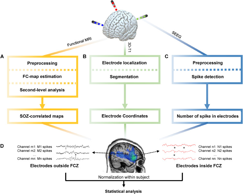
We studied 38 focal epilepsy patients who underwent simultaneous EEG/fMRI scanning and subsequent intracerebral stereo-EEG investigation. In EEG/fMRI analysis, IEDs with different spatial distributions were considered independent studies and IED-related maximal BOLD responses were evaluated. Studies with iEEG electrodes inside the maximal responses were selected and divided into three groups: Studies with 1. distinct maximal BOLD highly concordant with seizure-onset-zone (SOZ); 2. Moderate maximal BOLD concordant with SOZ; 3. maximal BOLD discordant with SOZ. Using maximal BOLD as seed, its functionally connected zone (FCZ) was determined. IED rates in iEEG channels inside and outside the FCZ were compared in the three groups. The effect of laterality and distance between channels and maximal BOLD, and correlation between functional connectivity values and IED rates were analyzed.
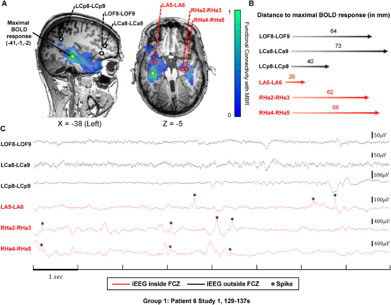
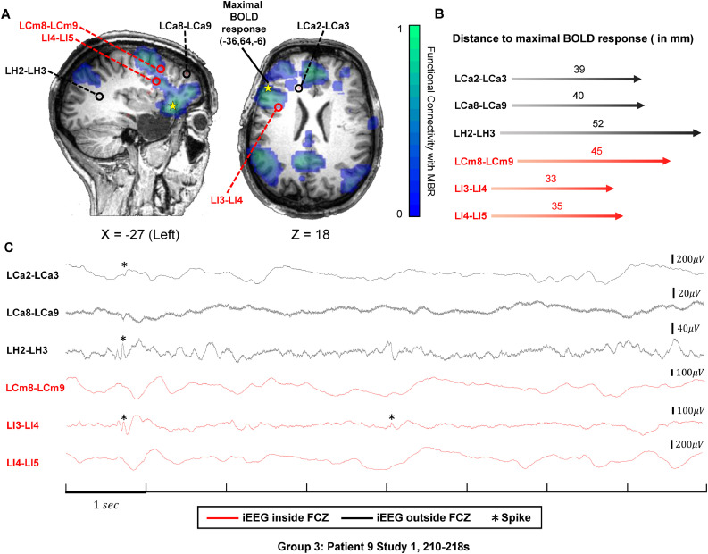
Thirty-six studies in 25 patients were included. IED rates of intracranial EEG channels inside the FCZ were significantly higher than outside in Group 1 (p = 2.6×10) and Group 2 (p = 1.2×10) and the inside-outside difference remained after regressing distance and laterality factors. In Group 1, connectivity values were significantly correlated with IED rates in channels inside the FCZ (p < 0.05).
Our results indicate a higher probability of finding intracranial IEDs in the FCZ of SOZ-concordant maximal BOLD responses than in other regions, regardless of distance and laterality. In studies with distinct maximal BOLD, connectivity values can partially predict IED rates in intracranial EEG. It is thus feasible to non-invasively delineate brain regions that are likely to have high IED rates.
探索颅内脑电图(iEEG)中不同脑区功能连接与发作间期癫痫放电(IEDs)存在的关系。
我们研究了 38 例接受同步脑电图/功能磁共振成像(fMRI)扫描和随后的颅内立体脑电图(EEG)研究的局灶性癫痫患者。在 EEG/fMRI 分析中,不同空间分布的 IEDs 被视为独立的研究,并评估了与 IED 相关的最大 BOLD 反应。选择有 iEEG 电极位于最大反应区内的研究,并将其分为三组:1. 与致痫区(SOZ)高度一致的明显最大 BOLD;2. 与 SOZ 一致的中度最大 BOLD;3. 与 SOZ 不一致的最大 BOLD。以最大 BOLD 为种子,确定其功能连接区(FCZ)。比较三组中 iEEG 通道内和 FCZ 外的 IED 率。分析通道与最大 BOLD 之间的侧别和距离的影响,以及功能连接值与 IED 率之间的相关性。
共纳入 25 例患者的 36 项研究。在组 1(p=2.6×10)和组 2(p=1.2×10)中,FCZ 内颅内 EEG 通道的 IED 率明显高于 FCZ 外,并且在回归距离和侧别因素后,内外差异仍然存在。在组 1 中,FCZ 内通道的连接值与 IED 率呈显著相关(p<0.05)。
我们的结果表明,与其他区域相比,SOZ 一致的最大 BOLD 反应的 FCZ 中更有可能发现颅内 IEDs,无论距离和侧别如何。在具有明显最大 BOLD 的研究中,连接值可以部分预测颅内 EEG 中的 IED 率。因此,无创描绘具有高 IED 率的脑区是可行的。