Bouatay Rachida, Aouf Lamia, Hmida Badii, El Korbi Amel, Kolsi Naourez, Harrathi Khaled, Koubaa Jamel
ENT Department at Fattouma Bourguiba Hospital, Monastir, Tunisie.
Radiology Department at Fattouma Bourguiba Hospital, Monastir, Tunisie.
Pan Afr Med J. 2019 Sep 2;34:3. doi: 10.11604/pamj.2019.34.3.18677. eCollection 2019.
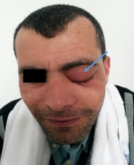
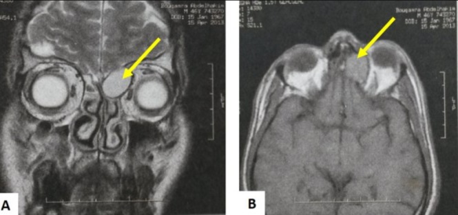
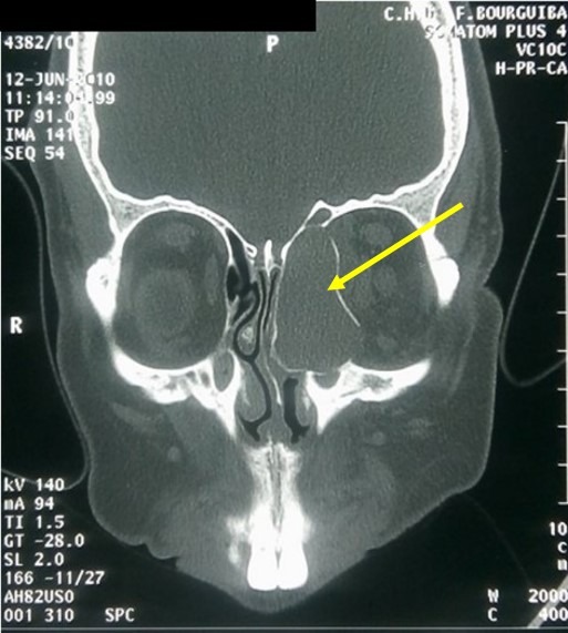
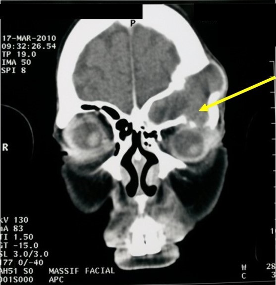
Mucoceles are slow-growing paranasal sinus cystic masses whose clinical presentation varies according to the affected sinus. Diagnosis is often radiological, based essentially on CT scan. The aim of this work was to study the radiologic characteristics of mucoceles on CT scan and MRI. We conducted a retrospective study of patients with mucoceles explored by imaging and operated on in our department. In our series, fronto-ethmoidal sinuses were the most frequently affected (81%). Facial scan confirmed the diagnosis in the majority of cases. Magnetic resonance imaging (MRI) was performed in 4 cases. Eleven patients were operated on by endonasal approach, three by external approach and one by combined surgical approach. Recurrence was observed in two patients after an average delay of 24 months. CT scan is considered the method of choice in the investigation of mucoceles. MRI is indicated in some cases to assess any orbital or intracranial extension.
黏液囊肿是生长缓慢的鼻窦囊性肿物,其临床表现因受累鼻窦而异。诊断通常依靠影像学检查,主要基于CT扫描。本研究的目的是探讨黏液囊肿在CT扫描和MRI上的影像学特征。我们对在我院接受影像学检查并手术治疗的黏液囊肿患者进行了一项回顾性研究。在我们的系列病例中,额筛窦最常受累(81%)。面部扫描在大多数病例中证实了诊断。4例患者进行了磁共振成像(MRI)检查。11例患者采用鼻内入路手术,3例采用外部入路手术,1例采用联合手术入路。2例患者在平均24个月的延迟后出现复发。CT扫描被认为是黏液囊肿检查的首选方法。在某些情况下,MRI用于评估眼眶或颅内有无扩展。