Department of Obstetrics and Gynaecology, The Second Xiangya Hospital, Central South University, 139 Renmin road, Changsha, 410011, Hunan, China.
Department of Obstetrics and Gynaecology, the First Affiliated Hospital of University of South China, Hengyang, 421001, Hunan, China.
BMC Cancer. 2019 Nov 28;19(1):1157. doi: 10.1186/s12885-019-6326-5.
Cervical cancer (CC), causing significant morbidity and mortality worldwide, is one of the most common gynecological malignancies in women. SFN has been reported as a potential prognostic marker with apparent high expression in tumors. Nevertheless, the function mechanism of SFN is not clear yet in CC.
The relative expressions of RNAs were detected by real-time quantitative PCR (RT-qPCR). Colony formation assay, EdU stained assay and CCK-8 assay were to check cell proliferation ability in CC. Flow cytometry and apoptosis related proteins analysis were used to measure cells apoptosis capacity. Luciferase reporter assay and RNA pull down assay were to verify the molecular mechanism.
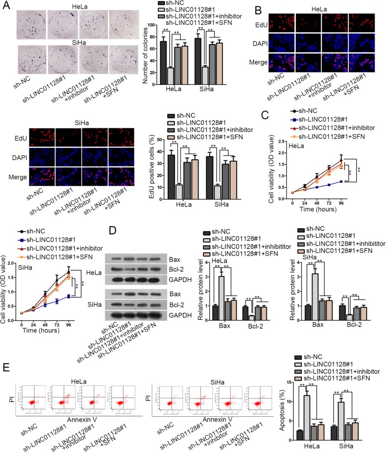
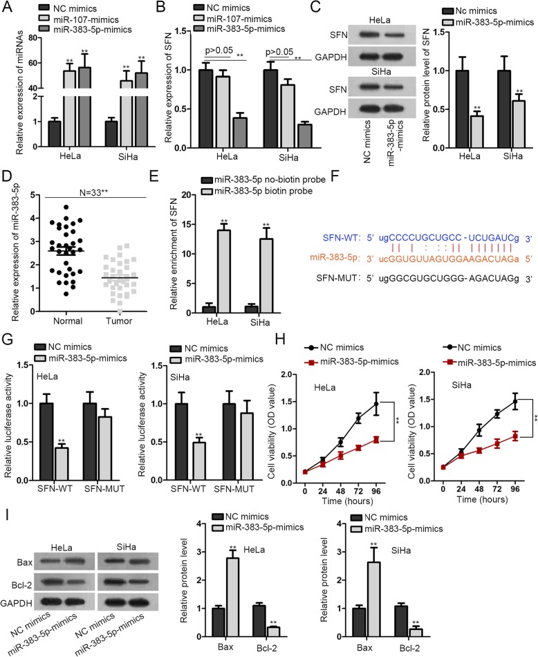
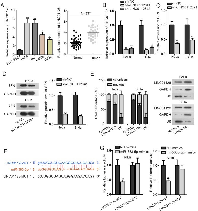
SFN was highly expressed in CC tissues and CC cell lines compared with normal tissues and normal cell line. After interfering SFN, cell proliferation, migration and invasion ability was inhibited as well as cell apoptosis ability was promoted. In subsequence, miR-383-5p exhibited conspicuous low expression in CC tissues. And miR-383-5p was found to bind to SFN and have anti-cancerous effects in CC. Moreover, LINC01128 displayed remarkable high expression in CC tissues. Besides, LINC01128 shortage could reduce the expression of SFN at mRNA and protein levels. And the affinity between LINC01128 and miR-383-5p was verified. In the end, it was proved that LINC01128 could enhance cell proliferation, migration and invasion as well as inhibit cell apoptosis by binding with miR-383-5p and upregulating SFN.
LINC01128 expedited cells cellular process in CC by binding with miR-383-5p to release SFN.
宫颈癌(CC)是一种常见的妇科恶性肿瘤,在全球范围内导致了相当高的发病率和死亡率。SFN 已被报道为一种潜在的预后标志物,在肿瘤中表达明显升高。然而,SFN 在 CC 中的作用机制尚不清楚。
采用实时定量 PCR(RT-qPCR)检测 RNA 的相对表达。集落形成实验、EdU 染色实验和 CCK-8 实验检测 CC 中细胞增殖能力。流式细胞术和凋亡相关蛋白分析检测细胞凋亡能力。荧光素酶报告基因实验和 RNA 下拉实验验证分子机制。
SFN 在 CC 组织和 CC 细胞系中的表达明显高于正常组织和正常细胞系。干扰 SFN 后,细胞增殖、迁移和侵袭能力受到抑制,细胞凋亡能力增强。随后,miR-383-5p 在 CC 组织中表达明显降低。miR-383-5p 与 SFN 结合,在 CC 中具有抗癌作用。此外,LINC01128 在 CC 组织中表达明显升高。此外,LINC01128 缺失可降低 SFN 的 mRNA 和蛋白水平表达。并验证了 LINC01128 与 miR-383-5p 的亲和力。最后,证明 LINC01128 通过与 miR-383-5p 结合释放 SFN,增强 CC 中的细胞增殖、迁移和侵袭,并抑制细胞凋亡。
LINC01128 通过与 miR-383-5p 结合释放 SFN,加速 CC 中的细胞进程。