Department of Neurosurgery, Nanjing Drum Tower Hospital Clinical College of Nanjing Medical University, Nanjing, 210008, Jiangsu, China.
Department of Neurosurgery, Nanjing Drum Tower Hospital, The Affiliated Hospital of Nanjing University Medical School, Nanjing, 210008, Jiangsu, China.
J Neuroinflammation. 2019 Nov 28;16(1):243. doi: 10.1186/s12974-019-1641-y.
Microglia are resident immune cells in the central nervous system and central to the innate immune system. Excessive activation of microglia after subarachnoid haemorrhage (SAH) contributes greatly to early brain injury, which is responsible for poor outcomes. Dehydroepiandrosterone (DHEA), a steroid hormone enriched in the brain, has recently been found to regulate microglial activation. The purpose of this study was to address the role of DHEA in SAH.
We used in vivo models of endovascular perforation and in vitro models of haemoglobin exposure to illustrate the effects of DHEA on microglia in SAH.
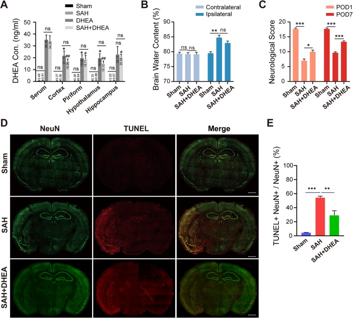
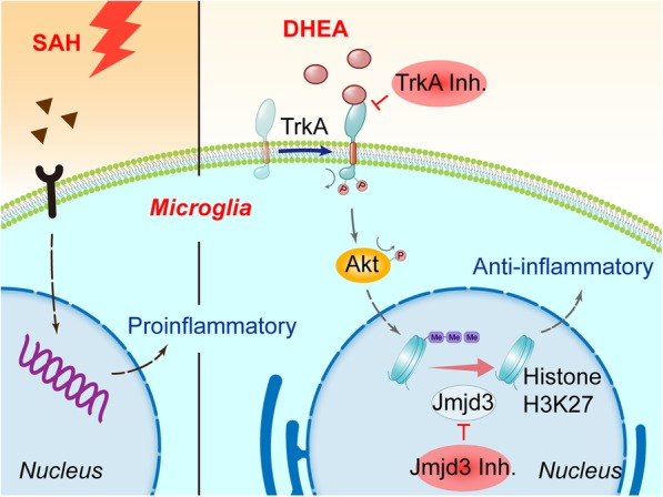
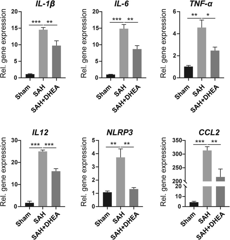
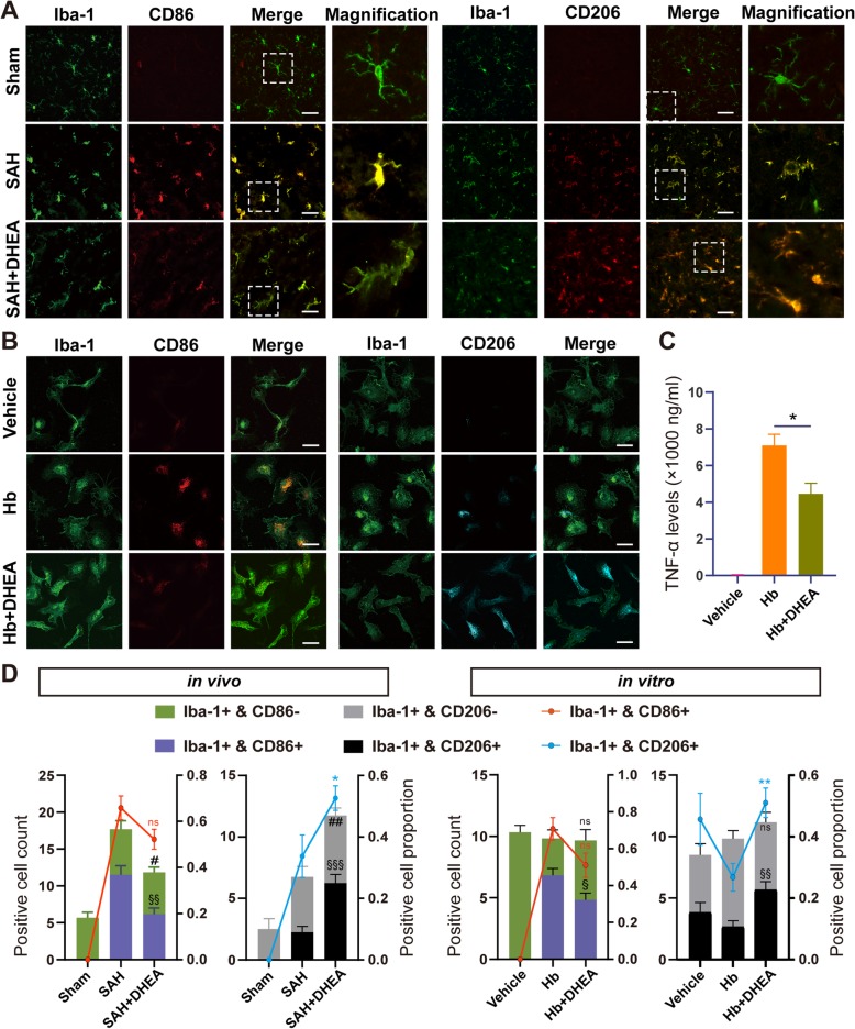
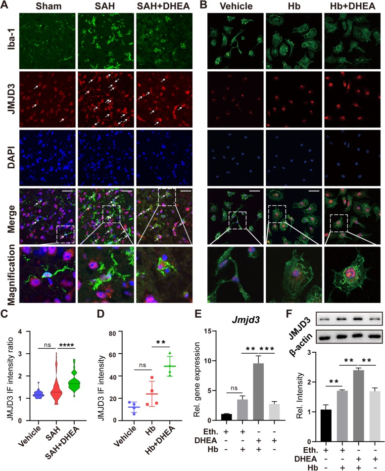
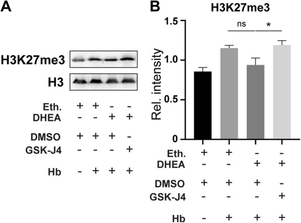
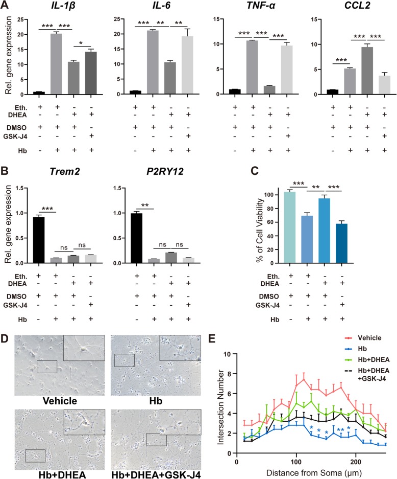
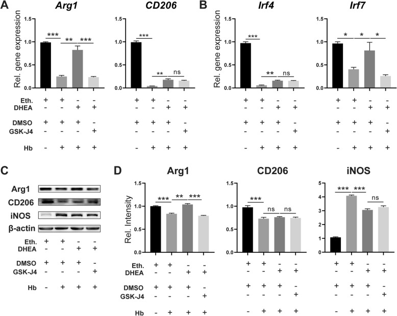
In experimental SAH mice, exogenous DHEA administration increased DHEA levels in the brain and modulated microglial activation. Ameliorated neuronal damage and improved neurological outcomes were also observed in the SAH mice pretreated with DHEA, suggesting neuronal protective effects of DHEA. In cultured microglia, DHEA elevated the mRNA and protein levels of Jumonji d3 (JMJD3, histone 3 demethylase) after haemoglobin exposure, downregulated the H3K27me3 level, and inhibited the transcription of proinflammatory genes. The devastating proinflammatory microglia-mediated effects on primary neurons were also attenuated by DHEA; however, specific inhibition of JMJD3 abolished the protective effects of DHEA. We next verified that DHEA-induced JMJD3 expression, at least in part, through the tropomyosin-related kinase A (TrkA)/Akt signalling pathway.
DHEA has a neuroprotective effect after SAH. Moreover, DHEA increases microglial JMJD3 expression to regulate proinflammatory/anti-inflammatory microglial activation after haemoglobin exposure, thereby suppressing inflammation.
小胶质细胞是中枢神经系统中的固有免疫细胞,是固有免疫系统的核心。蛛网膜下腔出血 (SAH) 后小胶质细胞过度激活极大地促成了早期脑损伤,这是导致预后不良的原因。脱氢表雄酮 (DHEA) 是一种富含大脑的类固醇激素,最近被发现可调节小胶质细胞的激活。本研究旨在探讨 DHEA 在 SAH 中的作用。
我们使用血管内穿孔的体内模型和血红蛋白暴露的体外模型来阐明 DHEA 对 SAH 中小胶质细胞的影响。
在实验性 SAH 小鼠中,外源性 DHEA 给药增加了大脑中的 DHEA 水平并调节了小胶质细胞的激活。在 DHEA 预处理的 SAH 小鼠中还观察到神经元损伤减轻和神经功能改善,这表明 DHEA 具有神经元保护作用。在培养的小胶质细胞中,DHEA 在血红蛋白暴露后上调了 Jumonji d3(JMJD3,组蛋白 3 去甲基化酶)的 mRNA 和蛋白水平,下调了 H3K27me3 水平,并抑制了促炎基因的转录。DHEA 还减弱了破坏性的促炎小胶质细胞对原代神经元的影响;然而,JMJD3 的特异性抑制消除了 DHEA 的保护作用。我们随后证实,DHEA 诱导的 JMJD3 表达至少部分是通过原肌球蛋白相关激酶 A(TrkA)/Akt 信号通路实现的。
DHEA 在 SAH 后具有神经保护作用。此外,DHEA 通过增加小胶质细胞中的 JMJD3 表达来调节血红蛋白暴露后的促炎/抗炎小胶质细胞激活,从而抑制炎症。