Zhang Juan, Yan Jifeng
Department of Cardiovascular Medicine, The Second Affiliated Hospital of Zhengzhou University, Zhengzhou, China.
Heart Center of Henan Provincial People's Hospital, Central China Fuwai Hospital, Zhengzhou, China.
Front Pharmacol. 2019 Nov 8;10:1241. doi: 10.3389/fphar.2019.01241. eCollection 2019.
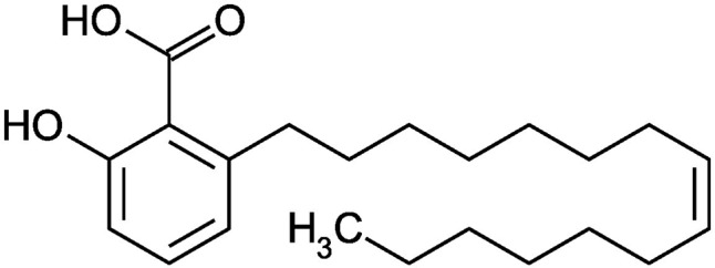
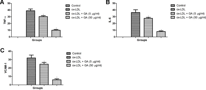
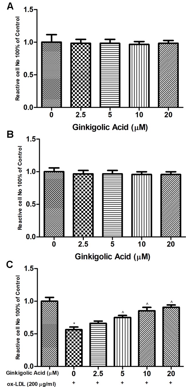
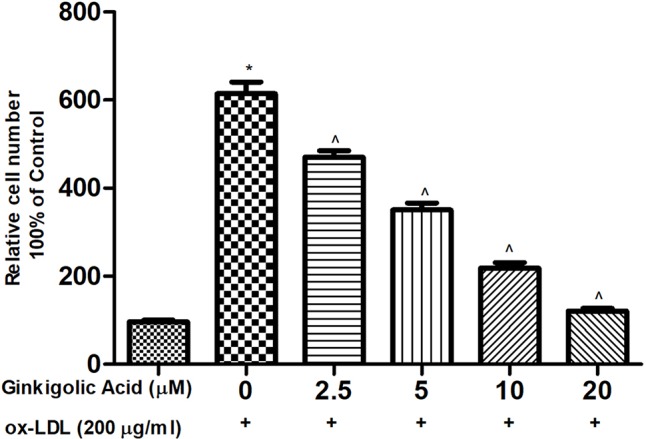
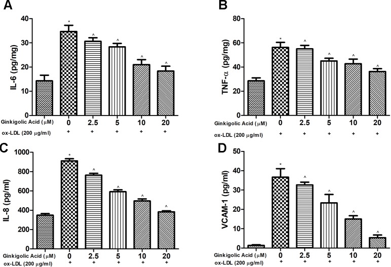
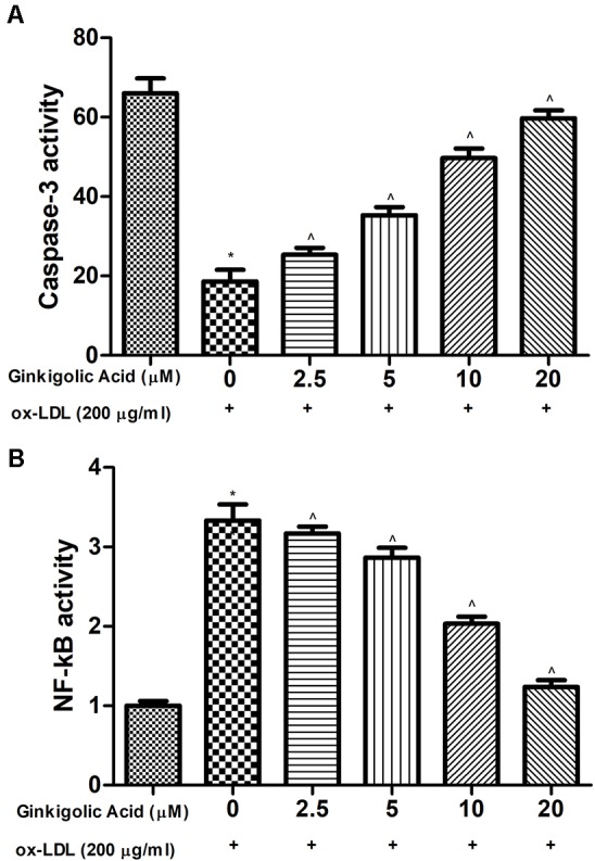
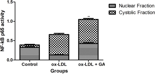
Oxidized low-density lipoprotein (ox-LDL) is considered as the significant maker of inflammatory reaction. ox-LDL was reported to play a crucial role in the pathogenesis of atherosclerosis (AS). In the current study, we scrutinize the suppressive effect of ginkgolic acid against ox-LDL induced an oxidative and inflammatory response in human microvascular endothelial cells (HMEC-1) and human peripheral blood mononuclear cells (nPBMCs) and explore the mechanism of action. HMEC-1 cells are treated with ox-LDL in the presence of different concentration of ginkgolic acid. MTT 3-(4,5-dimethylthiazol-2-yl)-2,5-diphenyltetrazolium bromide) assay was performed for the estimation of cell viability effect. Reactive oxygen species (ROS), inflammatory cytokines, and NF-κB activity are also estimated. For the hPBMCs assay, the cells were isolated from the healthy volunteers and cultured. The cells were further divided into different group and received the ginkgolic acid. Additionally, ROS, inflammatory marker such as prostaglandin E (PGE), lipoxygenase (LOX), nitric oxide (NO), cyclooxygenase (COX) protein expression, and mRNA expression of tumor necrosis factor-α (TNF-α), interleukin-6 (IL-6), and vascular cell adhesion protein 1 (VCAM-1) were estimated in the ox-LDL treated group. The result exhibited that ginkgolic acid treatment induced the cell viability boosting in ox-LDL treatment and intracellular ROS significantly decreased by ginkgolic acid. Pro-inflammatory cytokines also downregulated ginkgolic acid. Moreover, ginkgolic acid reduced the ox-LDL-induced NF-κB. The mRNA and protein expression of TNF-α, IL-6, and VCAM-1 considerably increased in the ox-LDL treated group and ginkgolic acid significantly reduced the mRNA and protein expression. An inflammatory marker such as PGE, LOX, and NO were increased in the ox-LDL treated group and ginkgolic acid treated group exhibited the reduction of an inflammatory marker. Based on the result, we can conclude that ginkgolic acid significantly reduced and reversed the ox-LDL-induced modulation, suggesting its anti-inflammatory effect the NF-κB pathway.
氧化型低密度脂蛋白(ox-LDL)被认为是炎症反应的重要标志物。据报道,ox-LDL在动脉粥样硬化(AS)的发病机制中起关键作用。在本研究中,我们仔细研究了银杏酸对ox-LDL诱导的人微血管内皮细胞(HMEC-1)和人外周血单核细胞(nPBMCs)氧化和炎症反应的抑制作用,并探讨其作用机制。在不同浓度银杏酸存在的情况下,用ox-LDL处理HMEC-1细胞。采用MTT(3-(4,5-二甲基噻唑-2-基)-2,5-二苯基四氮唑溴盐)法评估细胞活力效应。还对活性氧(ROS)、炎性细胞因子和核因子κB(NF-κB)活性进行了评估。对于人外周血单核细胞实验,从健康志愿者中分离细胞并进行培养。将细胞进一步分成不同组并给予银杏酸。此外,在ox-LDL处理组中评估了ROS、前列腺素E(PGE)、脂氧合酶(LOX)、一氧化氮(NO)等炎性标志物、环氧化酶(COX)蛋白表达以及肿瘤坏死因子-α(TNF-α)、白细胞介素-6(IL-6)和血管细胞黏附蛋白1(VCAM-1)的mRNA表达。结果显示,银杏酸处理可提高ox-LDL处理后的细胞活力,且银杏酸可显著降低细胞内ROS。银杏酸还下调了促炎细胞因子。此外,银杏酸降低了ox-LDL诱导的NF-κB。在ox-LDL处理组中,TNF-α、IL-6和VCAM-1的mRNA和蛋白表达显著增加,而银杏酸显著降低了其mRNA和蛋白表达。在ox-LDL处理组中,PGE、LOX和NO等炎性标志物增加,而银杏酸处理组则显示炎性标志物减少。基于这些结果,我们可以得出结论,银杏酸显著降低并逆转了ox-LDL诱导的调节作用,表明其通过NF-κB途径发挥抗炎作用。