Albert Einstein College of Medicine, 1300 Morris Park, Bronx, NY, 10461, USA.
Division of Plastic and Reconstructive Surgery, Keck School of Medicine of USC, 1510 San Pablo St. Suite 415, Los Angeles, CA, 90033, USA.
Sci Rep. 2019 Dec 4;9(1):18264. doi: 10.1038/s41598-019-54201-2.
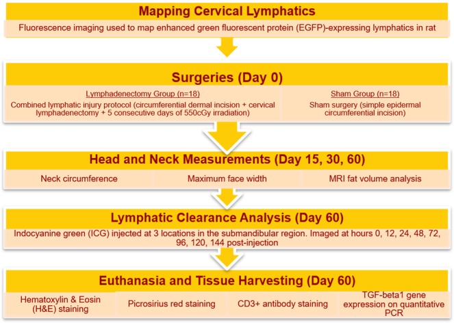
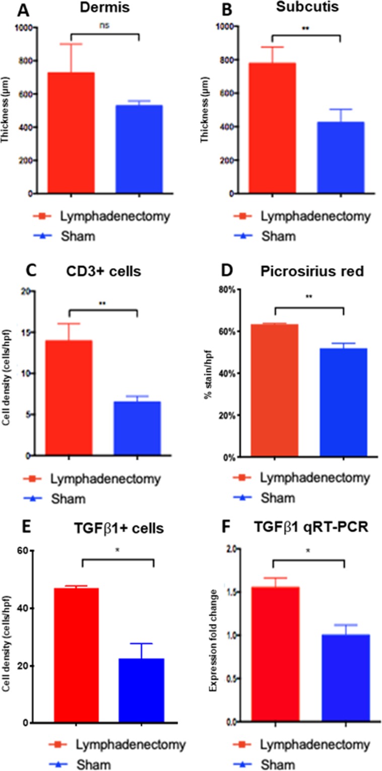
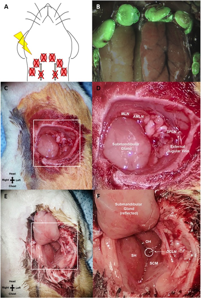
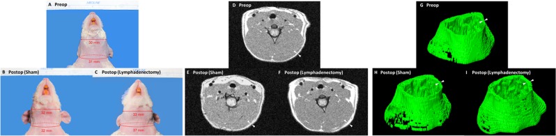
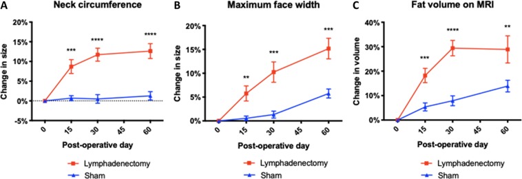
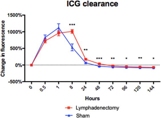
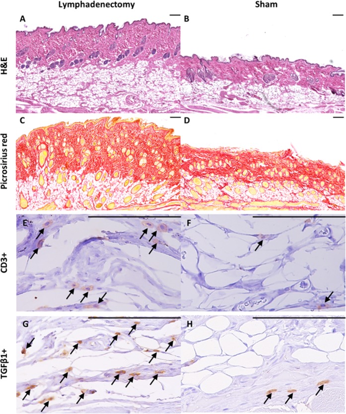
Head and neck lymphedema (HNL) is a disfiguring disease affecting over 90% of patients treated for head and neck cancer. Animal models of lymphedema are used to test pharmacologic and microsurgical therapies; however, no animal model for HNL is described in the literature to date. In this study we describe the first reproducible rat model for HNL. Animals were subjected to two surgical protocols: (1) lymphadenectomy plus irradiation; and (2) sham surgery and no irradiation. Head and neck expansion was measured on post-operative days 15, 30 and 60. Magnetic resonance imaging (MRI) was acquired at the same time points. Lymphatic drainage was measured at day 60 via indocyanine green (ICG) lymphography, after which animals were sacrificed for histological analysis. Postsurgical lymphedema was observed 100% of the time. Compared to sham-operated animals, lymphadenectomy animals experienced significantly more head and neck swelling at all timepoints (P < 0.01). Lymphadenectomy animals had significantly slower lymphatic drainage for 6 days post-ICG injection (P < 0.05). Histological analysis of lymphadenectomy animals revealed 83% greater subcutis thickness (P = 0.008), 22% greater collagen deposition (P = 0.001), 110% greater TGFβ1 cell density (P = 004), 1.7-fold increase in TGFβ1 mRNA expression (P = 0.03), and 114% greater T-cell infiltration (P = 0.005) compared to sham-operated animals. In conclusion, animals subjected to complete lymph node dissection and irradiation developed changes consistent with human clinical postsurgical HNL. This was evidenced by significant increase in all head and neck measurements, slower lymphatic drainage, subcutaneous tissue expansion, increased fibrosis, and increased inflammation compared to sham-operated animals.
头颈部淋巴水肿(HNL)是一种影响超过 90%头颈部癌症治疗患者的致残性疾病。淋巴水肿的动物模型用于测试药理和显微外科治疗;然而,迄今为止,文献中尚未描述用于 HNL 的动物模型。在这项研究中,我们描述了第一个可重现的 HNL 大鼠模型。动物接受了两种手术方案:(1)淋巴结切除术加放疗;(2)假手术和无放疗。术后第 15、30 和 60 天测量头颈部扩张。同时采集磁共振成像(MRI)。在第 60 天通过吲哚菁绿(ICG)淋巴管造影术测量淋巴引流,然后处死动物进行组织学分析。术后淋巴水肿 100%发生。与假手术动物相比,淋巴结切除术动物在所有时间点的头颈部肿胀明显更严重(P<0.01)。淋巴结切除术动物在 ICG 注射后 6 天的淋巴引流明显较慢(P<0.05)。淋巴结切除术动物的组织学分析显示,皮下组织厚度增加 83%(P=0.008),胶原沉积增加 22%(P=0.001),TGFβ1 细胞密度增加 110%(P=0.04),TGFβ1mRNA 表达增加 1.7 倍(P=0.03),T 细胞浸润增加 114%(P=0.005),与假手术动物相比。总之,接受完整淋巴结清扫和放疗的动物出现了与人类临床术后 HNL 一致的变化。与假手术动物相比,所有头颈部测量值均显著增加、淋巴引流缓慢、皮下组织扩张、纤维化增加和炎症增加证实了这一点。