Division of Plastic and Reconstructive Surgery, Department of Surgery, Memorial Sloan Kettering Cancer Center, New York, New York.
Division of Plastic and Reconstructive Surgery, Department of Surgery, Memorial Sloan Kettering Cancer Center, New York, New York.
Transl Res. 2019 Apr;206:57-70. doi: 10.1016/j.trsl.2018.12.003. Epub 2018 Dec 21.
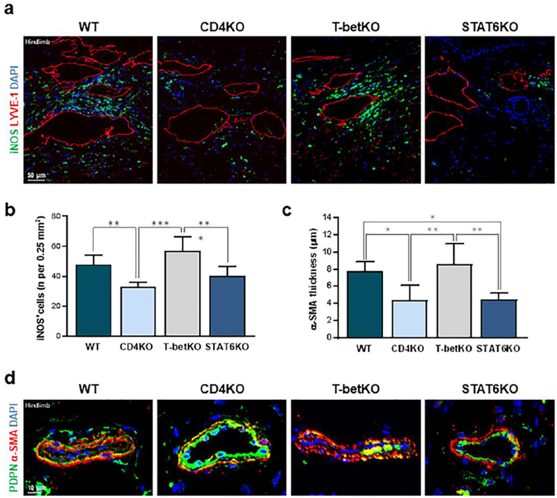
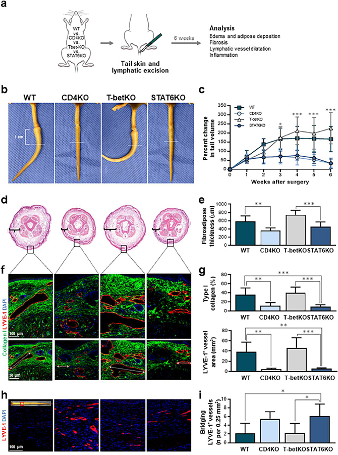
T cells infiltrating lymphedematous tissues have a mixed T helper 1 (Th1) and Th2 differentiation profile. Treatment with neutralizing antibodies targeting cytokines that promote Th2 differentiation (interleukin 4 [IL-4] and IL-13) decreases the severity of lymphedema in preclinical models, suggesting that Th2 cells play a key role in the pathology of this disease. However, these previous studies do not address the contribution of Th1 cells and it remains unknown if IL-4 and IL-3 blockade acts primarily on T cells or decreases the pathological changes of lymphedema by other mechanisms. Therefore, this study sought to analyze the effect of lymphatic injury in transgenic mice with mutations that cause defects in Th1 and Th2 cell generation (T-bet knockout or T-betKO and STAT6 knockout or STAT6KO mice, respectively). Using both the mouse tail and popliteal lymph node dissection models of lymphedema, we show that Th2-deficient (STAT6KO) mice are protected from developing lymphedema, have decreased fibrosis, increased collateral vessel formation, and preserved collecting lymphatic vessel pumping function. In contrast, mice with defective Th1 cell generation (T-betKO) develop disease with the same severity as wild-type controls. Taken together, our results suggest that Th2 differentiation is necessary for development of lymphedema following lymphatic injury and that Th1 differentiation does not significantly contribute to the pathology of the disease. Such findings are important as immunotherapy directed at Th2 cells has been found to be effective in well-studied Th2-mediated diseases such as asthma and atopic dermatitis and may therefore be similarly useful for lymphedema management.
浸润淋巴水肿组织的 T 细胞具有混合的 T 辅助 1(Th1)和 Th2 分化特征。用针对促进 Th2 分化的细胞因子(白细胞介素 4 [IL-4]和 IL-13)的中和抗体治疗可降低临床前模型中淋巴水肿的严重程度,表明 Th2 细胞在该疾病的病理学中起关键作用。然而,这些先前的研究并未解决 Th1 细胞的贡献问题,也不清楚 IL-4 和 IL-3 阻断是否主要作用于 T 细胞,还是通过其他机制减少淋巴水肿的病理变化。因此,本研究旨在分析导致 Th1 和 Th2 细胞生成缺陷的转基因小鼠(T 细胞因子盒结合蛋白 1 敲除或 T-betKO 和信号转导和转录激活因子 6 敲除或 STAT6KO 小鼠)的淋巴损伤的影响。使用小鼠尾巴和隐窝淋巴结解剖模型的淋巴水肿,我们表明 Th2 缺陷(STAT6KO)小鼠免受淋巴水肿的发展,纤维化减少,侧支血管形成增加,收集淋巴管泵功能得以保留。相比之下,Th1 细胞生成缺陷(T-betKO)的小鼠与野生型对照一样严重发病。总之,我们的结果表明,Th2 分化是淋巴损伤后淋巴水肿发展所必需的,而 Th1 分化对疾病的病理学没有显著贡献。这些发现很重要,因为针对 Th2 细胞的免疫疗法已被发现对研究充分的 Th2 介导疾病(如哮喘和特应性皮炎)有效,因此可能对淋巴水肿管理同样有用。