Cuciureanu Tudor, Huiban Laura, Chiriac Stefan, Singeap Ana-Maria, Danciu Mihai, Mihai Florin, Stanciu Carol, Trifan Anca, Vlad Nutu
Department of Gastroenterology, "Grigore T. Popa" University of Medicine and Pharmacy, "St. Spiridon" Emergency Hospital, Iasi 700115, Romania.
Department of Pathology, "Grigore T. Popa" University of Medicine and Pharmacy, "St. Spiridon" Emergency Hospital, Iasi 700115, Romania.
World J Clin Cases. 2019 Nov 26;7(22):3765-3771. doi: 10.12998/wjcc.v7.i22.3765.
Intestinal lipomas are rare benign gastrointestinal (GI) tumors, usually asymptomatic, but may become symptomatic as the result of some complications such as intussusception, intestinal obstruction, volvulus or bleeding. They can occur at any site along the entire GI tract, more frequent in colon and rarely in small intestine. The patient reported here is a very rare case of jejunal lipoma, ulcerated and intussuscepted, diagnosed in an adult investigated for a chronic iron deficiency anemia (IDA), and successfully managed by segmental jejunal resection.
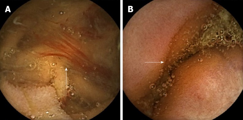
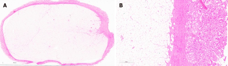
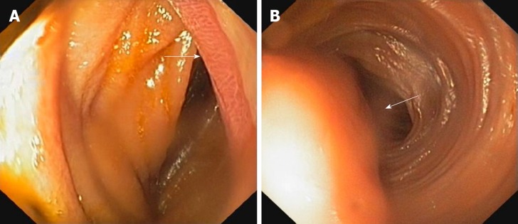
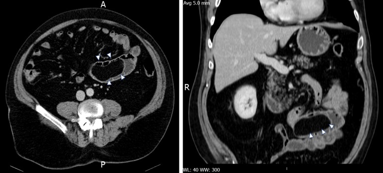
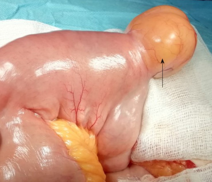
A 63-year-old male was referred to "St. Spiridon" Hospital, Institute of Gastroenterology and Hepatology, Iasi, to investigate an obscure GI bleeding with an IDA. After upper GI endoscopy and colonoscopy were performed, excluding potentially bleeding lesions, videocapsule endoscopy was then carried out, revealing fresh blood and a protruding lesion in proximal jejunum, findings confirmed by a single-balloon enteroscopy. Multiple biopsies were taken from the lesion, but histological results were inconclusive. Then, contrast - enhanced computed tomography was performed showing jejunal polypoid mass with homogenous fat density, suggestive for lipoma. A week later a laparotomy was performed revealing the intussuscepted jejunal segment which was resected , and sent for further histopathologic analysis. The patient made an uneventful recovery and was discharged seven days later, and at six months follow-up he had no complains and his hemoglobin returned to normal value.
Lipomas are very rarely located in the jejunum, usually asymptomatic, but they may lead to complications such as intussusception and bleeding. Surgical resection remains the treatment of choice.
肠道脂肪瘤是罕见的胃肠道良性肿瘤,通常无症状,但可能因肠套叠、肠梗阻、肠扭转或出血等并发症而出现症状。它们可发生于整个胃肠道的任何部位,在结肠更常见,在小肠则很少见。本文报道的患者是一例非常罕见的空肠脂肪瘤,伴有溃疡和肠套叠,在一名因慢性缺铁性贫血(IDA)接受检查的成年人中被诊断出来,并通过空肠节段切除成功治疗。
一名63岁男性因IDA伴不明原因的胃肠道出血被转诊至雅西胃肠病学和肝病研究所的“圣斯皮里东”医院。在进行了上消化道内镜检查和结肠镜检查,排除了潜在的出血病变后,进行了视频胶囊内镜检查,发现空肠近端有新鲜血液和一个突出病变,单气囊小肠镜检查证实了这些发现。从病变处取了多次活检,但组织学结果不明确。随后进行了对比增强计算机断层扫描,显示空肠息肉样肿块,密度均匀呈脂肪密度,提示为脂肪瘤。一周后进行了剖腹手术,发现了套叠空肠段并将其切除,送去做进一步的组织病理学分析。患者恢复顺利,7天后出院,6个月随访时无不适主诉,血红蛋白恢复正常。
脂肪瘤很少位于空肠,通常无症状,但可能导致肠套叠和出血等并发症。手术切除仍然是首选治疗方法。