Jakubowska Katarzyna, Koda Mariusz, Kisielewski Wojciech, Kańczuga-Koda Luiza, Famulski Waldemar
Department of Pathomorphology, Comprehensive Cancer Center, 15-027 Bialystok, Poland.
Department of General Pathomorphology, Medical University of Bialystok, 15-269 Bialystok, Poland.
Exp Ther Med. 2019 Dec;18(6):4904-4912. doi: 10.3892/etm.2019.8146. Epub 2019 Oct 30.
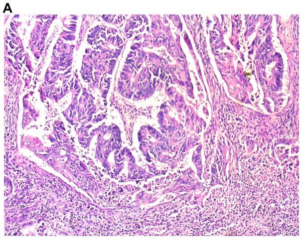
The presence of tumor cells in the large intestine stimulates hypoxia and local inflammatory mediators that activate numerous inflammatory cells, including a diverse lymphoid tumor cell population. The aim of the present study was to evaluate tumor-infiltrating lymphocytes (TILs) located in the invasive primary tumor, surrounding deposits of tumor cells and those present in distal metastatic cells in the liver of patients with colorectal cancer. Furthermore, the correlation of TILs with anatomical parameters was assessed. The study group included 123 patients with primary tumor colorectal cancer without distant metastasis, 25 cases with deposits of colorectal cancer cells and 15 cases of colorectal cancer liver metastasis. TILs were assessed in tissues stained with hematoxylin-eosin using light microscopy and evaluated by two independent pathologists blinded to the clinical information. Infiltration of TILs in the invasive front of primary tumor was stronger compared with those surrounding deposits of cancer cells and liver metastases (P<0.001). TILs in the invasive front of primary tumor masses were associated with various variables linked with tumor progression and inflammatory cell infiltrate. TILs distributed around the deposits of cancer cells were associated with postoperative treatment; however, those localized in the invasive front of liver metastases were correlated with preoperative therapy. In conclusion, TILs assessment in primary tumors of colorectal cancer, surrounding deposits of tumor cells and in the metastatic cells in the liver may be helpful in understanding the role of these cells in the organization of immune response.
大肠中肿瘤细胞的存在会引发缺氧和局部炎症介质,进而激活众多炎症细胞,包括多种淋巴样肿瘤细胞群体。本研究的目的是评估位于侵袭性原发性肿瘤、肿瘤细胞周围沉积物以及结直肠癌患者肝脏远端转移细胞中的肿瘤浸润淋巴细胞(TILs)。此外,还评估了TILs与解剖学参数的相关性。研究组包括123例无远处转移的原发性结直肠癌患者、25例结直肠癌细胞沉积物患者和15例结直肠癌肝转移患者。使用苏木精-伊红染色的组织通过光学显微镜评估TILs,并由两名对临床信息不知情的独立病理学家进行评估。原发性肿瘤侵袭前沿的TILs浸润比癌细胞周围沉积物和肝转移灶更强(P<0.001)。原发性肿瘤肿块侵袭前沿的TILs与多种与肿瘤进展和炎症细胞浸润相关的变量有关。癌细胞沉积物周围分布的TILs与术后治疗有关;然而,位于肝转移灶侵袭前沿的TILs与术前治疗相关。总之,评估结直肠癌原发性肿瘤、肿瘤细胞周围沉积物以及肝脏转移细胞中的TILs可能有助于理解这些细胞在免疫反应组织中的作用。