Department of Biomedical Engineering, Duke University, Durham, NC, 27710, USA.
School of Electrical and Computer Engineering, Cornell University, Ithaca, NY, 14853, USA.
Nat Commun. 2019 Dec 11;10(1):5647. doi: 10.1038/s41467-019-13699-w.
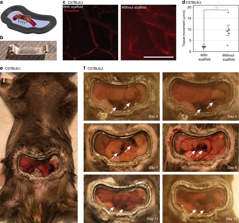
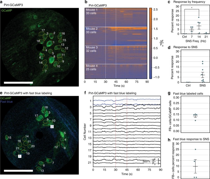
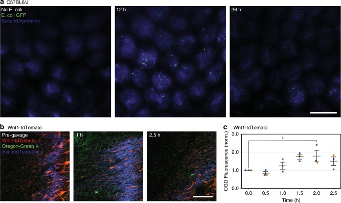
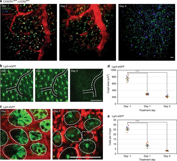
Intravital microscopy is a powerful technique to observe dynamic processes with single-cell resolution in live animals. No intravital window has been developed for imaging the colon due to its anatomic location and motility, although the colon is a key organ where the majority of microbiota reside and common diseases such as inflammatory bowel disease, functional gastrointestinal disorders, and colon cancer occur. Here we describe an intravital murine colonic window with a stabilizing ferromagnetic scaffold for chronic imaging, minimizing motion artifacts while maximizing long-term survival by preventing colonic obstruction. Using this setup, we image fluorescently-labeled stem cells, bacteria, and immune cells in live animal colons. Furthermore, we image nerve activity via calcium imaging in real time to demonstrate that electrical sacral nerve stimulation can activate colonic enteric neurons. The simple implantable apparatus enables visualization of live processes in the colon, which will open the window to a broad range of studies.
活体显微镜是一种强大的技术,可以在活体动物中以单细胞分辨率观察动态过程。由于结肠的解剖位置和运动性,目前还没有开发出用于结肠成像的活体窗口,尽管结肠是大多数微生物群所在的关键器官,并且常见疾病如炎症性肠病、功能性胃肠疾病和结肠癌也发生在这里。在这里,我们描述了一种带有稳定的铁磁性支架的活体鼠结肠窗口,用于慢性成像,通过防止结肠梗阻来最小化运动伪影,同时最大限度地提高长期存活率。使用这种设置,我们可以对活体动物结肠中的荧光标记的干细胞、细菌和免疫细胞进行成像。此外,我们通过钙成像实时成像神经活动,以证明电骶神经刺激可以激活结肠肠神经元。这个简单的可植入设备可以使人们直观地观察结肠中的活体过程,这将为广泛的研究开辟新的途径。