Giraldo-Villegas Manuela, Urquijo Jeaneth, Arnache-Olmos Oscar L, Rojas-López Mauricio
Grupo de Inmunología Celular e Inmunogenética, Sede de Investigación Universitaria (SIU), Universidad de Antioquia (UDEA), Calle 70 No. 52-21, Medellín, Colombia.
Grupo de Física del Estado Sólido, Sede de Investigación Universitaria (SIU), Universidad de Antioquia (UDEA), Medellín, Colombia.
Future Sci OA. 2019 Oct 30;5(10):FSO423. doi: 10.2144/fsoa-2019-0066.
To establish the effect of poly(acrylic acid)-coated iron oxide nanoparticles (PAC-IONs) and later exposure to a magnetic field on the differentiation of mononuclear phagocytes into macrophages.
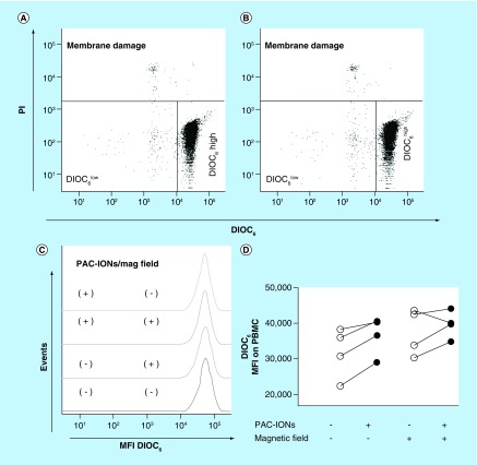
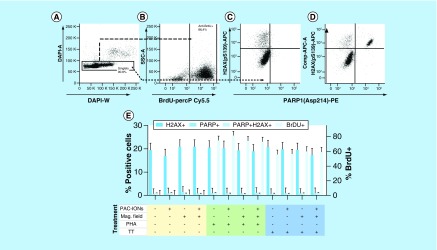
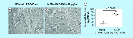
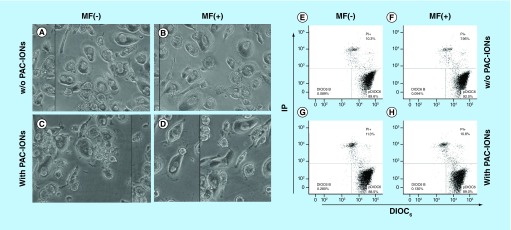
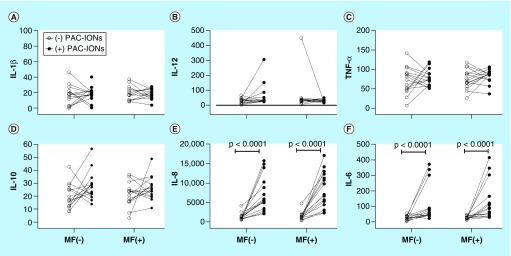
By flow cytometry, cell death was evaluated with DIOC6 and PI, Poly (ADP-ribose) Polymerases (PARP) fragmentation, H2AX phosphorylation and TUNEL assay. Cytokines by Cytokine bead array and the intracellular amount of iron by atomic absorption spectrometry.
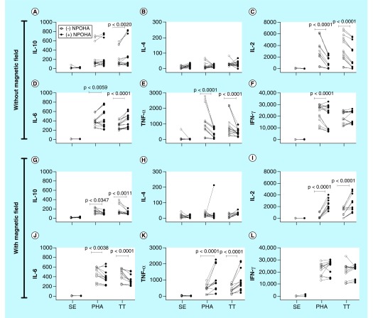
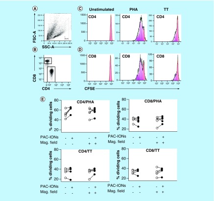
PAC-IONs did not induce apoptosis, modify the cell membrane integrity or alter the mitochondrial membrane potential. They did not affect the cell morphology, the pattern of cytokine accumulation or the activating role of differentiation of mononuclear phagocytes into macrophages on the proliferation of autologous T cells.
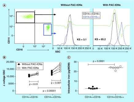
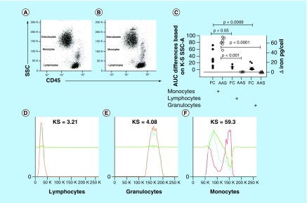
This evidence indicates that the PAC-IONs are safe and biocompatible. Moreover, the selectivity of the PAC-IONs for mononuclear phagocytes, as well as their increased uptake by non-classical monocytes, warrant future research with a view to their use as a contrast agent, a useful tool for in vivo tracking of tissue-infiltrating mononuclear phagocytes.
确定聚丙烯酸包被的氧化铁纳米颗粒(PAC-IONs)以及随后暴露于磁场对单核吞噬细胞分化为巨噬细胞的影响。
通过流式细胞术,使用DIOC6和PI、聚(ADP-核糖)聚合酶(PARP)片段化、H2AX磷酸化和TUNEL检测来评估细胞死亡。通过细胞因子珠阵列检测细胞因子,并通过原子吸收光谱法检测细胞内铁含量。
PAC-IONs未诱导细胞凋亡、改变细胞膜完整性或改变线粒体膜电位。它们不影响细胞形态、细胞因子积累模式或单核吞噬细胞分化为巨噬细胞对自体T细胞增殖的激活作用。
该证据表明PAC-IONs是安全且具有生物相容性的。此外,PAC-IONs对单核吞噬细胞的选择性以及它们被非经典单核细胞增加摄取的特性,值得未来开展研究以将其用作造影剂,这是一种用于体内追踪组织浸润单核吞噬细胞的有用工具。