Developmental Imaging and Biophysics Section, UCL Great Ormond Street Institute of Child Health, London, UK.
Sir Peter Mansfield Imaging Centre, School of Physics and Astronomy, University of Nottingham, Nottingham, UK.
MAGMA. 2020 Feb;33(1):141-161. doi: 10.1007/s10334-019-00800-z. Epub 2019 Dec 12.
This study aimed at developing technical recommendations for the acquisition, processing and analysis of renal ASL data in the human kidney at 1.5 T and 3 T field strengths that can promote standardization of renal perfusion measurements and facilitate the comparability of results across scanners and in multi-centre clinical studies.
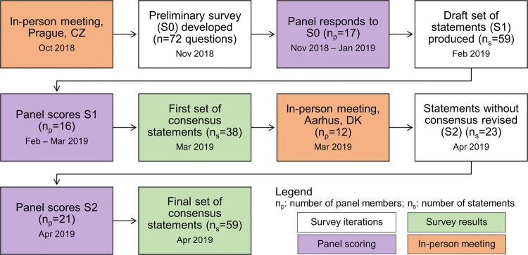
An international panel of 23 renal ASL experts followed a modified Delphi process, including on-line surveys and two in-person meetings, to formulate a series of consensus statements regarding patient preparation, hardware, acquisition protocol, analysis steps and data reporting.
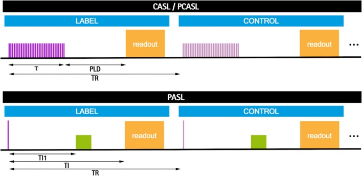
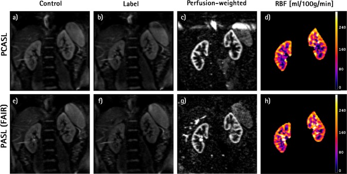
Fifty-nine statements achieved consensus, while agreement could not be reached on two statements related to patient preparation. As a default protocol, the panel recommends pseudo-continuous (PCASL) or flow-sensitive alternating inversion recovery (FAIR) labelling with a single-slice spin-echo EPI readout with background suppression and a simple but robust quantification model.
This approach is considered robust and reproducible and can provide renal perfusion images of adequate quality and SNR for most applications. If extended kidney coverage is desirable, a 2D multislice readout is recommended. These recommendations are based on current available evidence and expert opinion. Nonetheless they are expected to be updated as more data become available, since the renal ASL literature is rapidly expanding.
本研究旨在为 1.5T 和 3T 场强下人体肾脏 ASL 数据的采集、处理和分析制定技术建议,以促进肾灌注测量的标准化,并促进扫描仪间和多中心临床研究结果的可比性。
一个由 23 名肾脏 ASL 专家组成的国际小组采用改良 Delphi 流程,包括在线调查和两次现场会议,就患者准备、硬件、采集方案、分析步骤和数据报告制定了一系列共识声明。
59 项声明达成共识,而在与患者准备相关的两项声明上未能达成一致。作为默认协议,专家组建议使用单层面自旋回波 EPI 读取、背景抑制和简单但稳健的定量模型进行伪连续(PCASL)或流动敏感交替反转恢复(FAIR)标记。
这种方法被认为是稳健和可重复的,可以为大多数应用提供足够质量和 SNR 的肾灌注图像。如果需要扩展肾脏覆盖范围,则建议使用 2D 多层面读取。这些建议基于当前可用的证据和专家意见。然而,随着更多数据的出现,预计它们将被更新,因为肾脏 ASL 文献正在迅速扩展。