Laboratório de Vírus, Departamento de Microbiologia, Instituto de Ciências Biológicas, Universidade Federal de Minas Gerais, Belo Horizonte, Minas Gerais, 31270-901, Brazil.
Microbes, Evolution, Phylogeny and Infection (MEPHI), Aix-Marseille Université UM63, Institut de Recherche pour le Développement IRD 198, Assistance Publique-Hôpitaux de Marseille (AP-HM), Marseille, France.
Virol J. 2019 Dec 16;16(1):158. doi: 10.1186/s12985-019-1268-8.
After the isolation of Acanthamoeba polyphaga mimivirus (APMV), the study and search for new giant viruses has been intensified. Most giant viruses are associated with free-living amoebae of the genus Acanthamoeba; however other giant viruses have been isolated in Vermamoeba vermiformis, such as Faustovirus, Kaumoebavirus and Orpheovirus. These studies have considerably expanded our knowledge about the diversity, structure, genomics, and evolution of giant viruses. Until now, there has been only one Orpheovirus isolate, and many aspects of its life cycle remain to be elucidated.
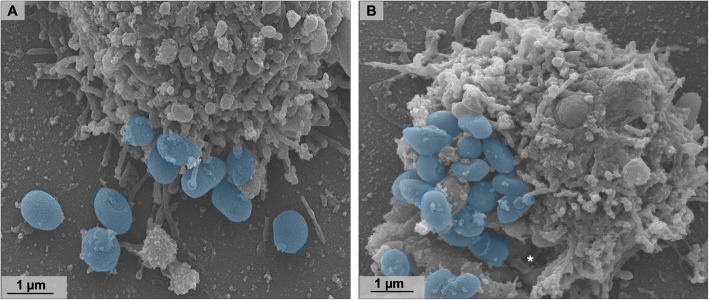
In this study, we performed an in-depth characterization of the replication cycle and particles of Orpheovirus by transmission and scanning electron microscopy, optical microscopy and IF assays.
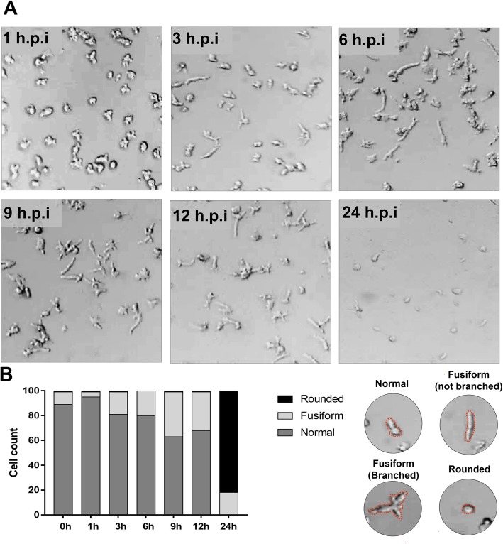
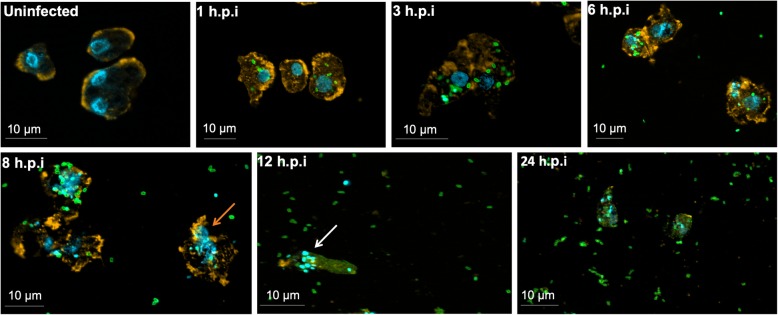
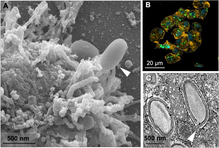
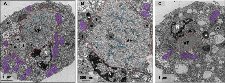
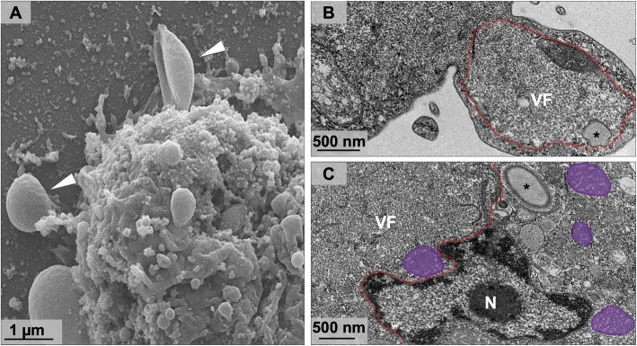
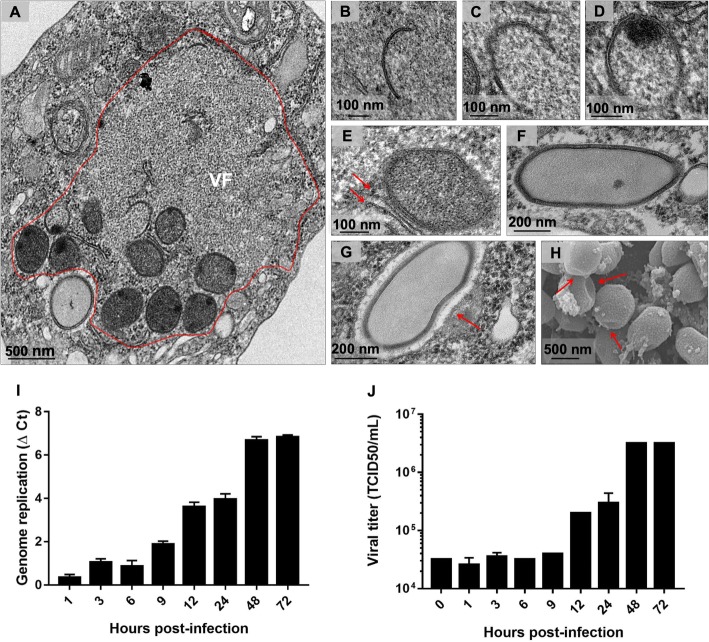
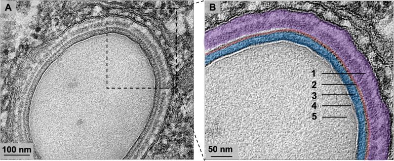
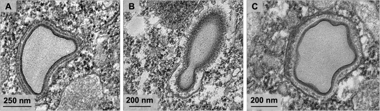
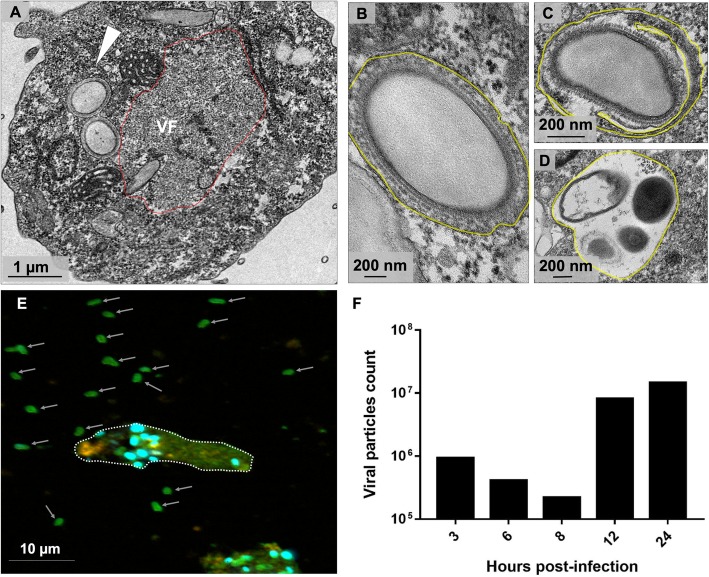
We observed, through optical and IF microscopy, morphological changes in V. vermiformis cells during Orpheovirus infection, as well as increased motility at 12 h post infection (h.p.i.). The viral factory formation and viral particle morphogenesis were analysed by transmission electron microscopy, revealing mitochondria and membrane recruitment into and around the electron-lucent viral factories. Membrane traffic inhibitor (Brefeldin A) negatively impacted particle morphogenesis. The first structure observed during particle morphogenesis was crescent-shaped bodies, which extend and are filled by the internal content until the formation of multi-layered mature particles. We also observed the formation of defective particles with different shapes and sizes. Virological assays revealed that viruses are released from the host by exocytosis at 12 h.p.i., which is associated with an increase of particle counts in the supernatant.
The results presented here contribute to a better understanding of the biology, structures and important steps in the replication cycle of Orpheovirus.
在分离出 Acanthamoeba polyphaga mimivirus(APMV)之后,对新型巨型病毒的研究和搜索得到了加强。大多数巨型病毒与自由生活的阿米巴属 Acanthamoeba 有关;然而,其他巨型病毒已在 Vermamoeba vermiformis 中分离出来,例如 Faustovirus、Kaumoebavirus 和 Orpheovirus。这些研究极大地扩展了我们对巨型病毒多样性、结构、基因组学和进化的认识。到目前为止,只有一种 Orpheovirus 分离株,其生命周期的许多方面仍有待阐明。
在这项研究中,我们通过透射和扫描电子显微镜、光学显微镜和 IF 测定法,对 Orpheovirus 的复制周期和颗粒进行了深入的表征。
通过光学和 IF 显微镜观察,我们观察到 V. vermiformis 细胞在 Orpheovirus 感染过程中的形态变化,以及感染后 12 小时(h.p.i.)时的运动性增加。通过透射电子显微镜分析了病毒工厂的形成和病毒颗粒形态发生,显示线粒体和膜募集到电子透明病毒工厂中及其周围。膜运输抑制剂(布雷菲德菌素 A)对颗粒形态发生产生负面影响。在颗粒形态发生过程中观察到的第一个结构是新月形体,其延伸并充满内部内容物,直到形成多层成熟颗粒。我们还观察到形成具有不同形状和大小的缺陷颗粒。病毒学测定法表明,病毒在 12 h.p.i. 通过胞吐作用从宿主中释放出来,这与上清液中颗粒计数的增加有关。
这里呈现的结果有助于更好地理解 Orpheovirus 的生物学、结构和复制周期中的重要步骤。