Department of Neurosurgery, The Second Affiliated Hospital of Soochow University, 1055, Sanxiang Road, Suzhou, 215004, China.
Department of Neurosurgery, the First Affiliated Hospital of Anhui Medical University, Hefei, 230022, China.
BMC Cancer. 2019 Dec 21;19(1):1240. doi: 10.1186/s12885-019-6460-0.
Tumor angiogenesis is vital for tumor growth. Recent evidence indicated that bone marrow-derived mesenchymal stem cells (BMSCs) can migrate to tumor sites and exert critical effects on tumor growth through direct and/or indirect interactions with tumor cells. However, the effect of BMSCs on tumor neovascularization has not been fully elucidated. This study aimed to investigate whether fusion cells from glioma stem cells and BMSCs participated in angiogenesis.
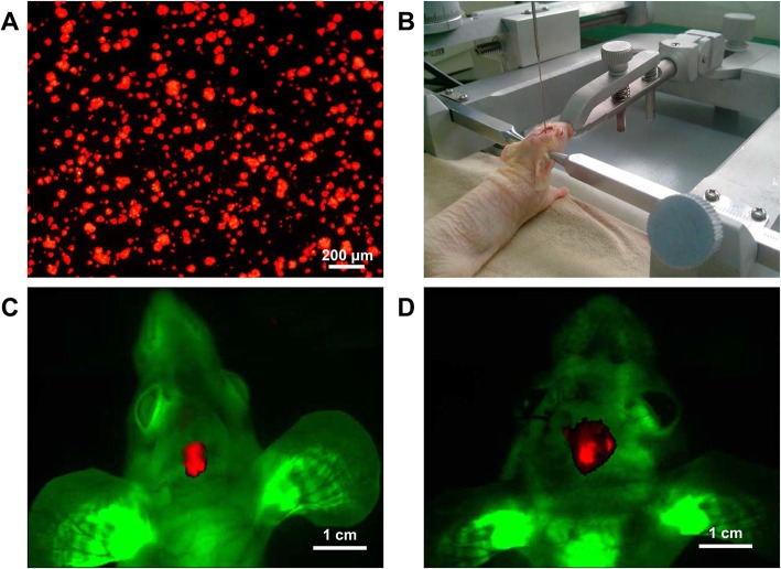
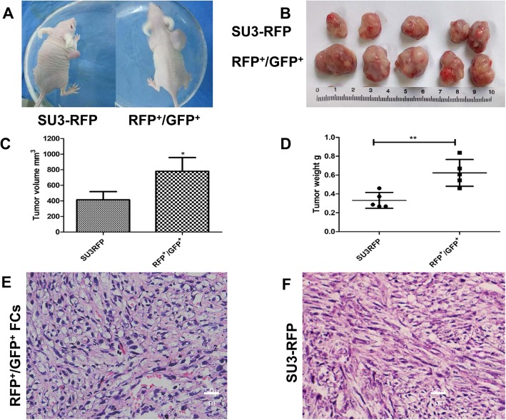
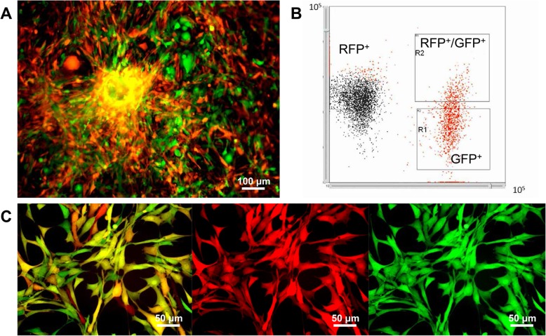
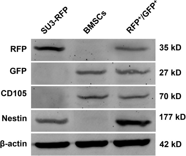
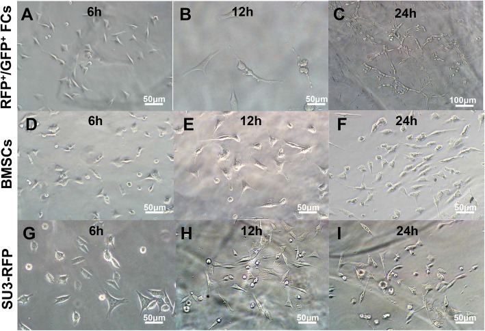
SU3-RFP cells were injected into the right caudate nucleus of NC-C57Bl/6 J-GFP nude mice, and the RFP+/GFP+ cells were isolated and named fusion cells. The angiogenic effects of SU3-RFP, BMSCs and fusion cells were compared in vivo and in vitro.
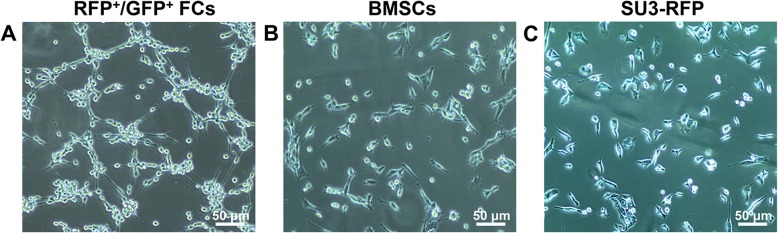
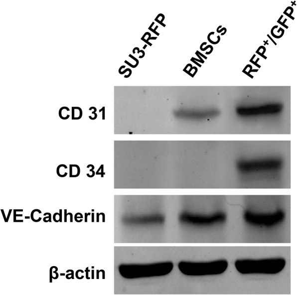
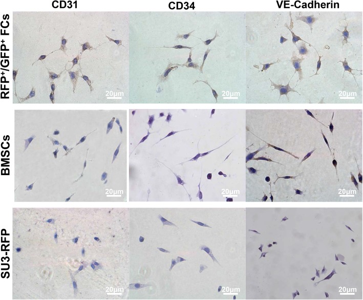
Fusion cells showed elevated levels of CD31, CD34 and VE-Cadherin (markers of VEC) as compared to SU3-RFP and BMSCs. The MVD-CD31 in RFP+/GFP+ cell xenograft tumor was significantly greater as compared to that in SU3-RFP xenograft tumor. In addition, the expression of CD133 and stem cell markers Nanog, Oct4 and Sox2 were increased in fusion cells as compared to the parental cells. Fusion cells exhibited enhanced angiogenic effect as compared to parental glioma cells in vivo and in vitro, which may be related to their stem cell properties.
Fusion cells exhibited enhanced angiogenic effect as compared to parental glioma cells in vivo and in vitro, which may be related to their stem cell properties. Hence, cell fusion may contribute to glioma angiogenesis.
肿瘤血管生成对于肿瘤生长至关重要。最近的证据表明,骨髓间充质干细胞(BMSCs)可以迁移到肿瘤部位,并通过与肿瘤细胞的直接和/或间接相互作用,对肿瘤生长产生关键影响。然而,BMSCs 对肿瘤新生血管形成的影响尚未完全阐明。本研究旨在探讨胶质瘤干细胞与 BMSCs 融合细胞是否参与了血管生成。
将 SU3-RFP 细胞注射到 NC-C57Bl/6-GFP 裸鼠右侧尾状核内,分离出 RFP+/GFP+细胞,并将其命名为融合细胞。在体内和体外比较了 SU3-RFP、BMSCs 和融合细胞的血管生成作用。
与 SU3-RFP 和 BMSCs 相比,融合细胞中 CD31、CD34 和 VE-Cadherin(VEC 标志物)的水平升高。RFP+/GFP+细胞异种移植瘤中的 MVD-CD31 明显高于 SU3-RFP 异种移植瘤。此外,与亲本细胞相比,融合细胞中 CD133 和干细胞标志物 Nanog、Oct4 和 Sox2 的表达增加。与亲本胶质瘤细胞相比,融合细胞在体内和体外均表现出增强的血管生成作用,这可能与其干细胞特性有关。
与亲本胶质瘤细胞相比,融合细胞在体内和体外均表现出增强的血管生成作用,这可能与其干细胞特性有关。因此,细胞融合可能有助于胶质瘤的血管生成。