Department of Ophthalmology, TOBB ETU School of Medicine, Ankara, Turkey
Clinic of Ophthalmology, Adıyaman University Training and Research Hospital, Adıyaman, Turkey
Balkan Med J. 2020 Apr 10;37(3):131-137. doi: 10.4274/balkanmedj.galenos.2020.2019.8.45. Epub 2020 Jan 7.
Corneal cross-linking treatment is the unique treatment method that can cease the progression of keratoconus disease. Because of the long duration of conventional treatment, accelerated cross-linking treatment methods are being developed.
To compare two different accelerated corneal cross-linking protocols in terms of postoperative visual acuity and topographic findings (higher-order aberrations and keratometry values).
Retrospective comparative study.
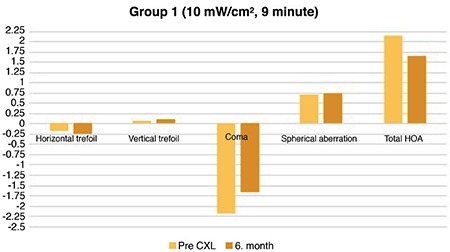
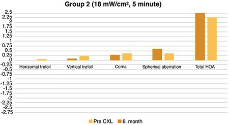
Sixty-five eyes of 43 patients (30 men and 13 women) who underwent two different accelerated corneal cross-linking protocols (10 min, 9 mW/cm and 5 min, 18 mW/cm) for progressive keratoconus were retrospectively analyzed. Patients were divided into two groups according to the accelerated corneal cross-linking treatment protocol: group 1 (10 min, 9 mW/cm, 32 eyes of 21 patients) and group 2 (5 min, 18 mW/cm, 33 eyes of 22 patients). Uncorrected visual acuity and best-corrected visual acuity values and topographic findings (central corneal thickness and flat and steep keratometry values) were recorded preoperatively and 6 months after corneal cross-linking treatment. High-order aberration values measured with Pentacam preoperatively and 6 months after corneal cross-linking were also recorded.
In both groups, a significant improvement was detected in the uncorrected visual acuity and best-corrected visual acuity levels preoperatively and 6 months postoperatively (group 1: p=0.001, p=0.001 and group 2: p=0.001, p=0.001, respectively). In addition, central corneal thickness values decreased significantly in both groups (p=0.006 and 0.001). Trefoil values showed no significant difference preoperatively and 6 months postoperatively in group 1 (p=0.160 and 0.620, respectively). In groups 1 and 2, coma values were found to decrease significantly in the 6 postoperative month compared with preoperative values (p=0.001 and 0.020, respectively). There was no significant difference between preoperative and 6 month postoperative horizontal and vertical trefoil values in both groups (p=0.850 and 0.140, respectively). There was no significant difference between the two groups in terms of preoperative and 6 month postoperative higher-order aberrations, refractive errors, keratometry values, and uncorrected visual acuity and best-corrected visual acuity levels.
Both accelerated corneal cross-linking procedures provide similar improvement in topographic findings, coma values and visual acuity.
角膜交联治疗是唯一能够阻止圆锥角膜进展的治疗方法。由于传统治疗时间较长,正在开发加速交联治疗方法。
比较两种不同的加速角膜交联方案在术后视力和地形学发现(高阶像差和角膜曲率值)方面的差异。
回顾性比较研究。
回顾性分析了 43 名患者(30 名男性和 13 名女性)的 65 只眼,这些患者因进展性圆锥角膜接受了两种不同的加速角膜交联方案(10 分钟,9 mW/cm 和 5 分钟,18 mW/cm)。根据加速角膜交联治疗方案将患者分为两组:第 1 组(10 分钟,9 mW/cm,32 只眼,21 名患者)和第 2 组(5 分钟,18 mW/cm,33 只眼,22 名患者)。记录术前和角膜交联治疗后 6 个月的未矫正视力和最佳矫正视力值以及地形学发现(中央角膜厚度和扁平及陡峭角膜曲率值)。术前和角膜交联治疗后 6 个月用 Pentacam 测量的高阶像差值也被记录下来。
两组患者术前和术后 6 个月的未矫正视力和最佳矫正视力水平均有显著提高(第 1 组:p=0.001,p=0.001;第 2 组:p=0.001,p=0.001)。此外,两组的中央角膜厚度值均显著降低(p=0.006 和 0.001)。第 1 组术前和术后 6 个月的三叶草值无显著差异(p=0.160 和 0.620)。第 1 组和第 2 组术后 6 个月的彗差值均显著低于术前值(p=0.001 和 0.020)。两组患者术后 6 个月的水平和垂直三叶草值与术前相比无显著差异(p=0.850 和 0.140)。两组患者在术前和术后 6 个月的高阶像差、屈光不正、角膜曲率值以及未矫正视力和最佳矫正视力水平方面均无显著差异。
两种加速角膜交联方案在地形学发现、彗差值和视力方面均提供了相似的改善。