From the A. A. Martinos Center for Biomedical Imaging, Department of Radiology, Massachusetts General Hospital, 149 13th St, Suite 2301, Charlestown, MA 02129 (X.L., B.S., B.T., O.C.A.); iCAD, Nashua, NH (K.J.); Departments of Neurosurgery (J.S., D.P.C.) and Neurology (J.D.), Massachusetts General Hospital, Boston, Mass; Department of Neurology, Brigham and Women's Hospital, Boston, Mass (T.T.B.); and Dana-Farber Cancer Institute, Boston, Mass (T.T.B.).
Radiology. 2020 Mar;294(3):589-597. doi: 10.1148/radiol.2020191529. Epub 2020 Jan 7.
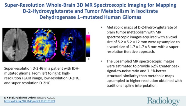
Background Isocitrate dehydrogenase (IDH) mutations are highly frequent in glioma, producing high levels of the oncometabolite D-2-hydroxyglutarate (D-2HG). Hence, D-2HG represents a valuable imaging marker for IDH-mutated human glioma. Purpose To develop and evaluate a super-resolution three-dimensional (3D) MR spectroscopic imaging strategy to map D-2HG and tumor metabolism in IDH-mutated human glioma. Materials and Methods Between March and September 2018, participants with IDH1-mutated gliomas and healthy participants were prospectively scanned with a 3-T whole-brain 3D MR spectroscopic imaging protocol optimized for D-2HG. The acquired D-2HG maps with a voxel size of 5.2 × 5.2 × 12 mm were upsampled to a voxel size of 1.7 × 1.7 × 3 mm using a super-resolution method that combined weighted total variation, feature-based nonlocal means, and high-spatial-resolution anatomic imaging priors. Validation with simulated healthy and patient data and phantom measurements was also performed. The Mann-Whitney test was used to check that the proposed super-resolution technique yields the highest peak signal-to-noise ratio and structural similarity index. Results Three participants with IDH1-mutated gliomas (mean age, 50 years ± 21 [standard deviation]; two men) and three healthy participants (mean age, 32 years ± 3; two men) were scanned. Twenty healthy participants (mean age, 33 years ± 5; 16 men) underwent a simulation of upsampled MR spectroscopic imaging. Super-resolution upsampling improved peak signal-to-noise ratio and structural similarity index by 62% ( < .05) and 7.3% ( < .05), respectively, for simulated data when compared with spline interpolation. Correspondingly, the proposed method significantly improved tissue contrast and structural information for the acquired 3D MR spectroscopic imaging data. Conclusion High-spatial-resolution whole-brain D-2-hydroxyglutarate imaging is possible in isocitrate dehydrogenase 1-mutated human glioma by using a super-resolution framework to upsample three-dimensional MR spectroscopic images acquired at lower resolution. © RSNA, 2020 See also the editorial by Huang and Lin in this issue.
背景 异柠檬酸脱氢酶 (IDH) 突变在胶质瘤中非常常见,产生高水平的致癌代谢物 D-2-羟戊二酸 (D-2HG)。因此,D-2HG 代表 IDH 突变型人类神经胶质瘤的一种有价值的成像标志物。 目的 开发和评估一种超高分辨率三维 (3D)MR 波谱成像策略,以绘制 IDH 突变型人类神经胶质瘤的 D-2HG 和肿瘤代谢图。 材料与方法 2018 年 3 月至 9 月,前瞻性纳入 IDH1 突变型胶质瘤患者和健康志愿者,使用优化的 3T 全脑 3D MR 波谱成像方案进行扫描,该方案针对 D-2HG 进行了优化。采用超高分辨率方法对采集的 D-2HG 图谱(体素大小为 5.2×5.2×12mm)进行上采样,体素大小为 1.7×1.7×3mm,该方法结合了加权全变差、基于特征的非局部均值和高空间分辨率解剖成像先验。还对模拟健康和患者数据以及体模测量值进行了验证。采用 Mann-Whitney U 检验检查所提出的超高分辨率技术是否能获得最高的峰值信噪比和结构相似性指数。 结果 共扫描了 3 例 IDH1 突变型胶质瘤患者(平均年龄,50 岁±21[标准差];2 例男性)和 3 例健康志愿者(平均年龄,32 岁±3;2 例男性)。20 例健康志愿者(平均年龄,33 岁±5;16 例男性)接受了模拟的上采样 MR 波谱成像。与样条插值相比,超高分辨率上采样可使模拟数据的峰值信噪比和结构相似性指数分别提高 62%(<.05)和 7.3%(<.05)。相应地,该方法可显著提高获取的 3D MR 波谱成像数据的组织对比度和结构信息。 结论 采用超高分辨率框架对上采样三维 MR 波谱图像可实现 IDH1 突变型人类神经胶质瘤的高空间分辨率全脑 D-2-羟戊二酸成像。