Laboratory of Animal Pathology and Public Health, Key Laboratory of Zoonosis of Ministry of Agriculture, College of Veterinary Medicine, China Agricultural University, Beijing, China.
Institute of Animal Husbandry and Veterinary Medicine, Beijing Academy of Agriculture and Forestry Sciences, Beijing, China.
Front Cell Infect Microbiol. 2019 Dec 20;9:433. doi: 10.3389/fcimb.2019.00433. eCollection 2019.
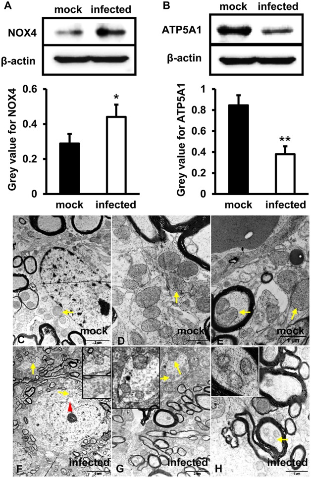
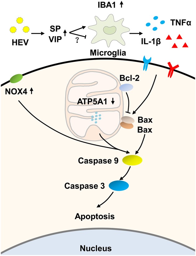
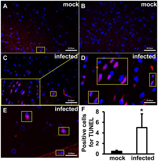
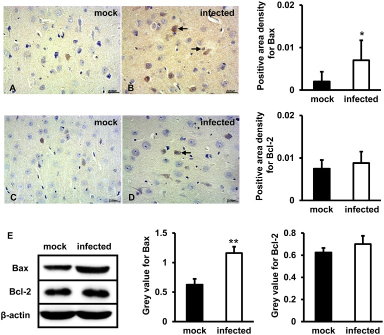
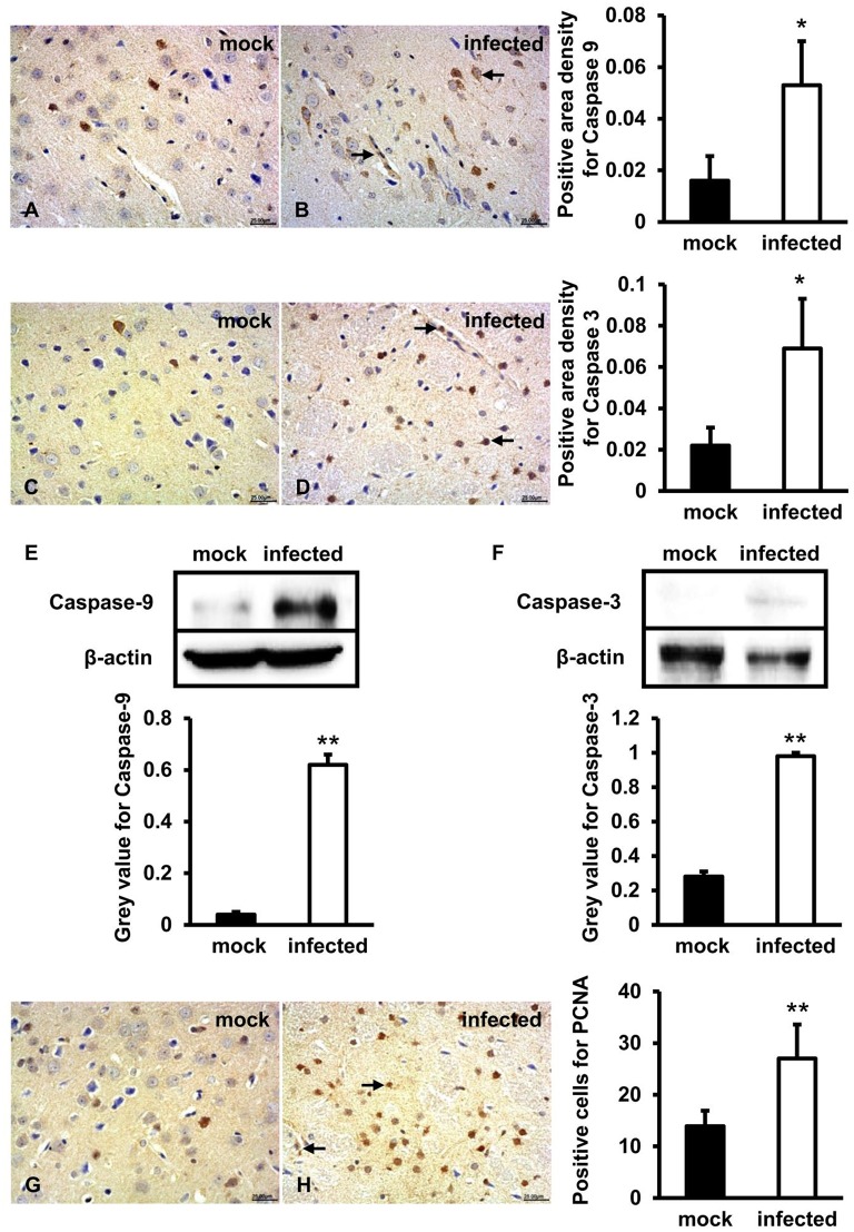
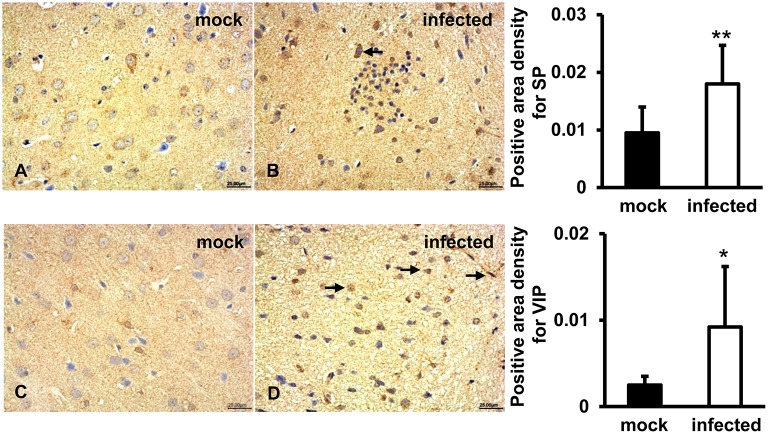
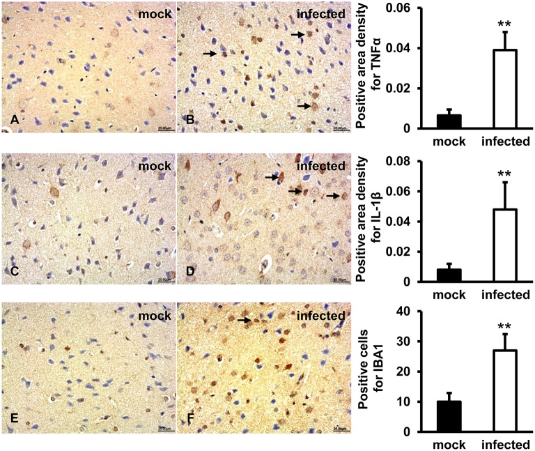
Hepatitis E virus (HEV) infection has been associated with extrahepatic manifestations, particularly neurological disorders. Although it has been reported that HEV infection induced hepatocyte apoptosis associated with mitochondria injury, activation of mitochondrial apoptotic pathway in the central nervous system during HEV infection was not clearly understood. In this study, the induction of mitochondrial apoptosis-associated proteins and pro-inflammatory cytokines were detected in HEV infected Mongolian gerbil model and primary human brain microvascular endothelial cells (HBMVECs). Mitochondrial exhibited fragments with loss of cristae and matrix in HEV infected brain tissue by transmission electron microscope (TEM). studies showed that expression of NADPH oxidase 4 (NOX4) was significantly increased in HEV infected HBMVECs ( < 0.05), while ATP5A1 was significantly decreased ( < 0.01). Expressions of pro-apoptotic proteins were further evaluated. Bax was significantly increased in both HEV infected brain tissues and HBMVECs ( < 0.01). studies showed that caspase-9 and caspase-3 were activated after HEV inoculation ( < 0.01), associated with PCNA overexpression as response to apoptosis. Cytokines were measured to evaluate tissue inflammatory levels. Results showed that the release of TNFα and IL-1β were significantly increased after HEV infection ( < 0.01), which might be attributed to microglia activation characterized by high level of IBA1 expression ( < 0.01). Taken together, these data support that HEV infection induces high levels of pro-inflammatory cytokines, associated with mitochondria-mediated apoptosis. The results provide new insight into mechanisms of extra-hepatic injury of HEV infection, especially in the central nervous system.
戊型肝炎病毒 (HEV) 感染与肝外表现有关,特别是神经紊乱。虽然有报道称 HEV 感染诱导与线粒体损伤相关的肝细胞凋亡,但在 HEV 感染期间中枢神经系统中线粒体凋亡途径的激活尚不清楚。在本研究中,我们检测了 HEV 感染蒙古沙鼠模型和原代人脑微血管内皮细胞 (HBMVEC) 中线粒体凋亡相关蛋白和促炎细胞因子的诱导。透射电镜 (TEM) 显示 HEV 感染脑组织中线粒体出现片段,嵴和基质丢失。研究表明,HEV 感染的 HBMVECs 中 NADPH 氧化酶 4 (NOX4) 的表达显著增加 ( < 0.05),而 ATP5A1 则显著降低 ( < 0.01)。进一步评估促凋亡蛋白的表达。Bax 在 HEV 感染的脑组织和 HBMVECs 中均显著增加 ( < 0.01)。研究表明,HEV 接种后 caspase-9 和 caspase-3 被激活 ( < 0.01),与 PCNA 过表达有关,以响应凋亡。测量细胞因子以评估组织炎症水平。结果表明,HEV 感染后 TNFα 和 IL-1β 的释放显著增加 ( < 0.01),这可能归因于小胶质细胞的激活,其特征是 IBA1 表达水平升高 ( < 0.01)。综上所述,这些数据支持 HEV 感染诱导高水平的促炎细胞因子,与线粒体介导的凋亡有关。这些结果为 HEV 感染的肝外损伤机制提供了新的见解,特别是在中枢神经系统。